Download
1 / 76
770 likes | 902 Views
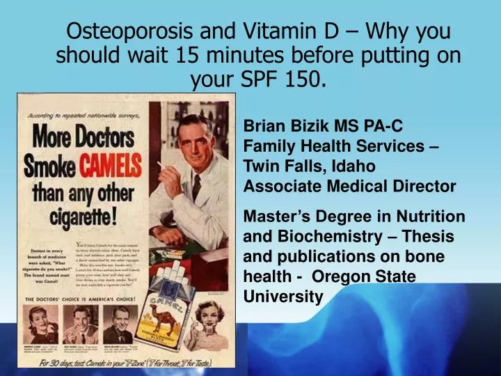
Osteoporosis and Vitamin D – Why you should wait 15 minutes before putting on your SPF 150. Brian Bizik MS PA-C Family Health Services – Twin Falls, Idaho Associate Medical Director

E N D
Osteoporosis and Vitamin D – Why you should wait 15 minutes before putting on your SPF 150. Brian Bizik MS PA-CFamily Health Services – Twin Falls, IdahoAssociate Medical Director Master’s Degree in Nutrition and Biochemistry – Thesis and publications on bone health - Oregon StateUniversity
WHAT WE ARE GOING TO COVER • The Disease itself • Diagnosis • Fragility Fractures • Treatment – when/how • FRAX
Big Picture Some general stuff – everyone has these slides so I do too.
Epidemiology – not much this won’t be on your boards. • A threat to 44 million or 55% of those age 50+ • 10 million (8 million women, 2 million men) have it, 34 million have low bone mass. • In 2005 there were 297,000 hip fractures; 547,000 vertebral fractures; 397,000 wrist fractures and 135,000 pelvic fractures. • BUT – most fractures happen in women with LOW bone mass: -1.0 to -2.4. Hmmmm. . .
2 cases • 76 Y O female – falls, breaks hip – spends 4 months in skilled nursing facility – never recovers and long term placement is recommended. Could this have been prevented? • 66 Y O female with a radial fracture after getting up off the floor while playing with her grandson. What does this mean?
Definition of osteoporosis “…a systemic skeletal disease characterized by low bone mass and micro-architectural deterioration of bone tissue, leading to enhanced bone fragility and a consequent increase in fracture risk.” World Health Organization (WHO), 1994
Big Picture Lets talk about bone in general This is it!!
TYPES OF BONE (1) Cortical – is hard, compact, dense bone (example: long-bones) (2) Trabecular – is spongy, porous and flexible bone (example: end of thewrist, hip and the spine). Much more sensitive to hormonal changes.
HEALTHY BONE Bone is living tissue, which is constantly being broken down and rebuilt, a process called remodeling Bone is renewed all the time – 15% is actively being changed at any given time
OSTEOPOROTIC BONE The loss of bony matrix leading to weakened bones – the point at which fracture becomes a real threat.
BONE “REMODELING” Osteoclasts – dig channels – resorb bone - WHY???? Osteoblasts – lay down new matrix (protein and mineral)
Big Picture Okay – what is happening to bones AND what is a fragility fracture?
Age-related changes in femoral neck cortex and association with hip fracture Mayhew et al, Lancet 2005 20-year-old 80-year-old Those with hip fractures have: • Preferential thinning of the inferior anterior cortex • Increased cortical porosity
Age-related changes in bone properties associated with fracture risk • Decreased bone mass and BMD • Altered geometry • Altered architecture • Cortical thinning • Cortical porosity • Trabecular deterioration young elderly
Tibia Radius Premenopausal Postmenopausal Osteopaenia Postmenopausal Osteoporosis Postmenopausal Severe Osteoporosis
Osteoporotic fragility fractures This is the key, wake up for just a minute and learn this, then resume your post-Disney nap. Some key points before we continue:
Osteoporotic fragility fracturesaka – grandma should not break a bone from a ground level fall Hip Shoulder Knee Elbow
Incidence of fractures Only 30% of vertebral fractures are “clinically apparent” 700,000 750,000 500,000 Annual incidence 300,000+ 300,000+ 200,000 250,000 Clinically apparent 0 Vertebral Hip Wrist Other Fracture type
250,000 / year 700,000 / year Over 300,000 / year Other sites: 300,000 / year
Distal radius fracture • The most common fracture in women at middle age • Incidence increase begins just after menopause in women • The most common fracture in men below age 70 years Holmberg et al, Osteoporos Int. 2006; 7:1065-77
Proximal humerus fracture • Estimated that fractures of the proximal part of the humerus account for 4-8 % of all fractures • In persons over 40, fractures of the proximal humerus account for 76% of all fractures of the humerus • Data suggest that fracture of the proximal humerus is the third most common fracture over age 65 • Fractures of the proximal humerus have shown a pattern of increase similar to other common fragility fractures
Pathogenesis of fragility fractures Neuromuscular function Environmental risks Age Type of fall Energy reduction External protection Bone mass Bone structure Bone quality Fall Risk Impact of fall Skeletal strength Fracture risk
Height Loss/Steroids • Any height loss over 1.5 inches needs to be thought of as an osteoporotic fracture (many microfractures). • Steroid therapy is a big risk factor. 5mg of prednisone or equivalent for 3 or more months is a risk factor.
Bone Mass vs. Age Heaney RP et al: Peak bone mass. Osteo Int 2000;11:985.
Big Picture What is happening to old people?
4000 3500 Females 3000 Males 2500 2000 1500 1000 500 0 0-4 5-14 15-24 25-34 35-44 45-54 55-64 65-74 75-84 85+ Age- and sex-specific incidence of all limb fractures Incidence per 100,000 person-years Age
Men Hip Vertebrae Forearm 35 55 75 Osteoporotic fracture incidence Women 4,000 3,000 Hip Incidence per 100,000 person-years 2,000 Vertebrae 1,000 Forearm 35 55 75 Age
10000 10000 8000 8000 6000 6000 4000 4000 2000 2000 0 0 Whole bone strength declines dramatically with age Femoral neck (sideways fall) Lumbar vertebrae (compression) 20 y Whole bone strength (Newtons) 70 y 20 y old
Prevalence of osteoporosis in women at different skeletal sites 60 T-score ≤ -2.5 50 40 Prevalence (%) 30 20 10 0 50-59 60-69 70-79 80+ age 50+ Age (years) Spine Hip Mid-radius Any site Melton et al. J Bone Miner Res 1995; 10:175
Prevalence of osteoporosis at the femoral neck in Caucasian women 50% 40% Prevalence 30% 20% 10% 0% 50-54 55-59 60-64 65-69 70-74 75-79 80-84 Age (years)
Outcome after hip fracture The situation one year after fracture
3 cell types work together to remodel bone OSTEOCYTE BONE MATRIX
Bone remodeling occurs throughout life Remodeling
Big Picture A 3 minute review of your last nutrition class
Review of the Nutrients - Calcium • Calcium – needed for bone mineralization • Key is to build bone early – peak in the early 20’s!! • Once peak is reached, preserve bone with adequate calcium – 1200-1500 mg per day (impossible without dairy or supplements) • Each serving about 300 mg – supplements don’t have much calcium
Review of the Nutrients - Calcium • Women’s Health Initiative – overall, those with calcium supp. had no decrease in fractures – but when just those who complied with pill taking were separated out – there was a stat. sig. drop in fractures. NNT with Ca was 5000, NNT with Ca and Vit D was 3000. • Tell your patients that “calcium, and vitamin D are necessary but not sufficient”. • If they drink pop (esp. if instead of milk), the added phosphorus will dramatically increase the amount of calcium lost in the urine.
Review of the Nutrients part deux • Vitamin D – the new “in” test. MANY are very low – often very low. Ideal levels above 30 ng/ml. I guar-en-tee if you test 10 chronic pain patients you will get 9 that are below 30 ng/ml, many below 15 ng/ml which means severely deficient. HUGE role with muscle strength and coordination (fall risk). • D2 is Ergocalciferol. Not natural. Made from yeast or other. Found in many supplements, it is cheaper. • D2 is 1/7th as potent as D3. It is converted to D3 readily unless you have CKD – anyone with a GFR under 40 may not convert D2 to D3 at all.
Review of the Nutrients part tre’ If you give supplemental D2, provide an Rx for 50,000 units of D2 or Ergocalciferol qweek for 8 weeks then recheck. • D3 is the natural form in our blood. We make this from cholesterol and sunlight. Foods, as a source of D3, really stink (ie – fish and other stuff not found at Taco Bell) • Sunlight works below Sacramento or so year round and all over US in Summer (one study in Boston showed zero Vit D3 Nov – Feb). But, you have to get it. Sunscreen blocks it all (UVB). 10-15 minutes gives more than you can ever get from food. • Serum tests look at D3. So if you give D2, and they don’t convert D2 to D3, it won’t do much. Give D3!
Review of the Nutrients part quad D3 supplementation works very well for everyone. It is relatively cheap. Give Calcitrol (Rocaltrol) 0.25 to 0.50 mcg PO QD. Give daily for 4-8 weeks then if levels normalized then switch to daily D2 Pearl - If giving bisphosphonate and no improvement in BMD in 2 years consider this as the failure cause. Pearl 2 – any D3 level below 30 ng/ml will cause a rise in parathyroid hormone (remember that one. . Serum calcium falls, PTH goes up, calcium absorption in gut goes up, urinary excretion goes down, bone remineralization goes up, serum calcium goes back up, PTH goes back down to normal)
Big Picture OK, who is at risk. . . Come on. . . who should I test??
Risk Factors • This is key – this is who to send for DEXA and this is how you know who to treat. • Age, Gender, history of fracture for patient or family. • Thin, use of steroids (more than 3 months), other bone issues (RA) • Smoking and excessive ETOH. • AND. . . BMD!
Bone Mineral Density (BMD) • Again – just one risk factor, but the most important one. • Analyzed with a DEXA scan. • Send any patient age 50+ with multiple risk factors -or- US Preventative Services Task Force recommends all women over 65 and men over 70 should be screened. • Send anyone over 50 with ANY minor trauma that causes a fracture. – like a ground level fall. Many of these are fragility fractures. Some data that traumatic fractures are an indication for screening as well - even if MVA. But if sure a FF, then DEXA not needed to decide if you should treat. Maybe good for monitoring effect.
Bone Mineral Density (BMD) • Check BMD on patients you are either monitoring or treating as often as every two years. You may not need it that frequently. • Results given in T-score and Z-score for both hip and spine. • Z-score will compare grandma to grandma (compare to age matched controls). Only useful if doing a DEXA on a young person or if you suspect something other than primary osteoporosis. • T-score will compare grandma to a young white woman. (BMD vs. young healthy bone) WHO uses this one. So you use this one. This is the one we base BMD determinations on.
WHO criteria for osteoporosis T-score: Remember – this is the difference expressed as standard deviation compared to young white reference population
Osteoporotic fracture and BMDThere is no magic number! Fractures per 1,000 person-years Number of fractures 50 Fracture rate Women with fractures 400 40 300 30 200 20 100 10 0 0 1.0 0.5 0.0 -0.5 -1.0 -1.5 -2.0 -2.5 -3.0 -3.5 Siris et al. Arch Intern Med. 2004; 164:1108-1112
DEXA and Bone –some real numbers! • Consider testing q2y in women who are near a treatment threshold either by DEXA or FRAX. • Over testing – if they have no other risk factors and BMD not likely to be low enough to treat, don’t test. But how do you know? • For women with osteopenia on DEXA, there was an annual rate of bone loss is about 0.5% of BMD per year which is about 0.1 point loss of T-score • No data that repeat testing improves outcome. • In one large study, 73% of women on therapy who lost BMD at year 1 showed a gain by year 2.
Diagnosing osteoporosis DEXA (dual-energy x-ray absorptiometry)
Any other reason (other than being old) to have low BMD? In one study 15% of women with Celiac Disease presented with osteoporosis as their first sign/symptom. Other conditions to consider is Cushing’s Syndrome, alcoholism, CHF and depression/diet changes as conditions that may spur osteoporosis.
To treat or not to treat Let’s look at the treatment options first, then roll all this up into one huge ball of phlegm.