Download
1 / 61
610 likes | 896 Views
Cutaneous Fungal Infections. DR. NOUF TALAL MILEH DERMATOLOGY DEMONSTRATOR. Cutaneous Fungal Infections. Dermatomycosis - general name for any skin disease caused by a fungus.

E N D
Cutaneous Fungal Infections DR. NOUF TALAL MILEH DERMATOLOGY DEMONSTRATOR
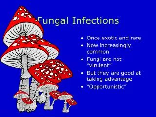
Cutaneous Fungal Infections • Dermatomycosis - general name for any skin disease caused by a fungus. • Dermatophytosis - "ringworm" disease of the nails, hair, and/or stratum corneum of the skin caused by fungi called dermatophytes.
Cutaneous Fungal Infections • Etiological agents are called dermatophytes . • Three important anamorphic genera are involved in ringworm. 1)Microsporum. 2)Trichophyton. 3) Epidermophyton.
Cutaneous Fungal Infections • Dermatophytes are keratinophilic- "keratin loving". Digest keratin by their keratinases • Keratin is a major protein found in horns, hooves, nails, hair, and skin. • Ringworm - disease called ‘herpes' by the Greeks, and by the Romans ‘tinea' (which means small insect larvae).
DERMATOPHYTES • Classified into three groups depending on their usual habitat ANTROPOPHILIC keratin-utilizing on hosts - humans (e.g., M. audounii, T. tonsurans, Trichophytonrubrum ) GEOPHILIC keratin-utilizing soil saprophytes (e.g., M. gypseum…). ZOOPHILIC keratin-utilizing on hosts - living animals (e.g., M. canis, T. verrucosum).
DERMATOPHYTOSISPathogenesis and Immunity • Contact and trauma. • Moisture. • Crowded living conditions. • Cellular immunodeficiency (chronic inf.). • Re-infection is possible (but, larger inoculum is needed, the course is shorter ).
Major sources of ringworm infection • Schools, military camps, prisons. • Warm damp areas (e.g., tropics, moisture accumulation in clothing and shoes). • Animals (e.g., dogs, cats, cattle, poultry, etc.). • Historical note: More people were shipped out of the Pacific Theater in WWII back to U.S. because of ringworm infection then through injury.
Transmission • Close human contact. • Sharing clothes, combs, brushes, towels, bedsheets... (fomites). • Animal-to-human contact (Zoophilic).
DERMATOPHYTOSIS Clinical Classification Infection is named according to the anatomic location involved: • Tineacapitis- ringworm infection of the head, scalp, eyebrows, eyelashes. • Tineafacialis- of the face. • Tineacorporis- of the body . • Tineacruris- of the groin (jock itch). • Tineaunguium- of the nails. • Tineabarbae- of the beard. • Tineamanuum- of the hand. • Tineapedis- of the foot (athlete's foot).
CLINICAL MANIFESTATIONS OF RINGWORMTineapedis - Athletes' foot infection • Between toes or toe webs (releasing of clear fluid) - 4th and 5th toes are most common, sole . • Commonly, patients describe pruritic, scaly soles and, often, painful fissures between the toes. • Most frequently due to: - Trichophyton (T.) rubrum - T. interdigitale, previouslycalledT. mentagrophytes var. interdigitale - Epidermophytonfloccosum
Tineapedis - Athletes' foot infection • 3 possible clinical presentations: ( Interdigital, Chronichyperkeratotic (moccasin) Inflammatory or vesiculobullous ) • Allergic reactions are sometimes associated with tineapedis and other ringworm infections.
Tineacorporis - body ringworm • Generally restricted to stratum corneumof the smooth skin. • Symptoms result form fungi metabolites such as toxin/allergens. • Produces concentric or ring-like lesions on skin (annular plaques), and in severe cases these are raised and may become inflamed.
Tineacorporis - body ringworm • All forms of tineacorporis caused by T. rubrum, T. mentagrophytes, T. tonsurans, M. canis, and M. audouinii are treatable with topical agent containing ketoconazole, miconazole, etc... • Widespread tineacorporis and more severe types (lesions) require systemic griseofulvintreatment (about 6 weeks for effective treatment).
Tineacruris - ringworm of the groin and surrounding region • More common in men than women. • Infection seen on scrotum and inner thigh, the penis is usually not infected. • Several causes of tineacruris include T. rubrum , E. flocossum. • Predisposing factors include persistent perspiration, high humidity, tight clothing , diabetes and obesity, topical glucocorticoid application.
Tineacruris - ringworm of the groin and surrounding region • Symptom: none, prurits. • Signs: large, scaling well demarcated red / tan/ brown/ plaques. • Diagnosis • If lesion "weep", it is likely caused by a yeast, such as, Candida albicans, and not by a dermatophyte, especially if infections are seen in a woman.
Tineaunguium - ringworm of the nails Tineaunguium or onychomycosis can take 4 forms : • Distal subungualonychomycosis • Proximal subungualonychomycosis • Superficial white onychomycosis • Candidalonychomycosis
Tineaunguium - ringworm of the nails • Most commonly caused by T. rubrum, then E. floccosumor other Trichophyton species. • Resistant to treatment, rarely resolves spontaneously.
Tineaunguium - ringworm of the nails • Topical treatments - poor record of cure. • If the disease involve one or two nails loceryl nail loquer ( amorolfiene ). • If the disease involve multable nails lamfine (terbinafine) 250 mg (6 w for fingure nail, 12 w for toe nail ) • Systemic griseofulvin therapy can lead to remission .
Tineafacialis • Dermatophytosis of the glabrous facial skin. • More common in children. • Etiology : T. tonsurans ,T. rubrum, T. mentagrophytes , M. audouinii, M. canis . • Predisposing factors : animal exposure, topical application of glucocorticoid. • Symptoms : asymptomatic • Characterized by a well-circumscribed erythematous patch, minimal scaling.
TineaBarbae • Involving the beard and moustache areas. • Males only, adult. • Etiology : T. verrucosum, T. mentagrophytes. • Predisposing factors : more common in farmers. • Symptoms : pruritus, tenderness, pain. • Signs : scattered, discrete follicular pustules and papules. • D.D. : Beard folliculitis ( s.aures ) • Treatment : see tineacapitis.
Tineacapitis - ringworm of the scalp, eyebrows and eyelashes • Predominantly a disease of preadolescent children • Caused by species of Microsporumand Trichophyton. • Fungus grows into hair follicle. • Using a Wood's lamp, on hair Microsporum species tend to fluoresce green while Trichophyton species generally do not fluoresce. • Lack of fluorescence does not mean it isn't Microsporum.
Tineacapitis • classified according to the microscopic pattern of fungal invasion as endothrix, ectothrix, and favus. Endothrix : • Fungi invade the inside of the hair shaft. • composed of fungal arthroconidie and hyphae, without cuticle destruction. • The causative organisms TVS.
Tineacapitis Ectothrix : • Fungi invade the outside of the hair shfat. • The process of ectothrixinvasion is similar to endothrix invasion, with the exception that the hyphae destroy the hair cuticle, thenconvert intoinfectiousarthroconidia. • The causative organisms are M. audouinii, M. canis, M distortum, T. ferrugineum. All theseorganismscause fluorescence under Wood light. ( to remamber, SEE CATS AND DOGS FIGHT)
Tineacapitis Favus • Is a type of inflammatory tinea that is characterized by invasion of hair by hyphyae not produce conidia, and presence of linear air spaces. • Mainly caused by T schoenleinii.
Clinical Manifestations • Endothrix ( black dot ) • Ectothrix ( gray patch ) • Kerion • favus
TineaCapitisClinical Manifestations • Clinical Characteristics according to Causative Agent
Diagnosis • ClinicalDiagnosis. • Direct Examination: ( hyphae and arthrospores ) • Culture : (Mycobiotic agar ,Sabouraud dextrose agar )
Ectothrix and Endothrix Fluorescing hair (under Wood's lamp)
Diagnosis • ImmunologicStudy: ( type I response and a delayed type IV response ) • Histopathology : ( growth of hyphae and formation of arthroconidia )
Rapid identification and differentiation of fungal DNAin dermatological specimens by LightCycler PCRRalf Gutzmer, Susanne Mommert, Uta Ku¨ ttler, Thomas Werfeland Alexander Kapp • In conclusion, we developed a fast and simple LightCyclerbased PCR to detect DNA of the dermatologically most relevant fungi and to differentiate dermatophytes, yeasts and non-dermatophyte moulds. • This can complement mycological culture and direct microscopy in the diagnosis of fungal skin disease and provides additional diagnostic information in a substantial number of patients. Journal of Medical Microbiology (2004), 53, 1207–1214ormation in a substantial number of patients
Differential Diagnosis According to Clinical Presentation • Diffuse scaling (noninflammatory) : Seborrheic and atopic dermatitis, psoriasis. • Alopecia plaque (noninflammatory) :Seborrheic and atopicdermatitis, psoriasis. • Black dots : Alopeciaareata, trichotillomania. • Diffuse pustular(inflammatory) : Bacterialfolliculitis • kerioncelsi(inflammatory): Abscess, tumor.
Treatment TopicalTreatment : • Shampoocontaining 2% seleniumsulfide. • shampooscontaining 2% ketoconazole. • Topical antifungal cream : ketoconazole (nezoral), micinazole ( daktarin)
Oral Griseofulvin( treatment of choice ) • Fungistatic, and inhibits nucleic acid synthesis, arresting cell division at metaphase and impairing fungal cell wall synthesis. • It is also anti-inflammatory • Tablet or suspension form • Recommended dose: for pediatric microsized15 mg/kg/day, ultramicrosized 10 mg/kg/day • For adults up to 250 mg/ day, Bid.
Griseofulvin • between 8 and 12 weeks. • Side effects nausea and rashes in 8-15%. • contra-indicated in pregnancy, lupus erythematosus, porphyria and severe liver disease. • Advantages inexpensive, effective, safe. • Disadvantages: Prolonged treatment required. • Drug interactions :warfarin, cyclosporin and the oral contraceptive pill.
Terbinafine • Acts on fungal cell membranes and is fungicidal. • It is effective against all dermatophytes • Effective as griseofulvin and is safe for the management of scalp ringworm due to Trichophyton sp. • Dosage : 250 mg qd, depends on the weight of the patient ( if the weight 10-20 kg 62.5 mg, between 20-40 kg 125 mg, > 40 kg 250 mg.
Terbinafine • Side-effects include; gastrointestinal disturbances and rashes in 5% and 3% of cases, • Advantages. Fungicidal so shorter therapy required (cf. griseofulvin) so increased compliance more likely. • Disadvantages. No suspension formulation .
Itraconazole • exhibits both fungistatic and fungicidal activity • Fungistatic through depletion of cell membrane ergosterol, which interferes with membrane permeability. • Doses in children 5 mg/kg per day. • in adult 200 mg/day. • Duration 4 weeks.
Itraconazole • Advantage. Pulsed shorter treatment regimens are possible. • Disadvantage. possible side-effects. Potential drug interactions. • Drug interactions. Enhanced toxicity of anticoagulants (warfarin), antihistamines (terfenadine and astemizole), antipsychotics (sertindole), anxiolytics (midazolam), digoxin.
Fluconazole • Doses : for pediatric 6 mg/kg per day for 4 weeks for adult : 200 mg/ day Ketoconazole • Doses : for pediatric 5 mg/kg per day for adult 200-400 mg / day its use in children is limited by hepatotoxicity
Terbinafinehydrochloride oral granules versus oralgriseofulvin suspension in children with tineacapitis:Results of two randomized, investigator-blinded,multicenter, international, controlled trials* • Objective: To compare the efficacy and safety of a new pediatric formulation of terbinafine hydrochloride oral granules with griseofulvin oral suspension in the treatment of tineacapitis. • Results:Rates of complete cure and mycologic cure were significantly higher for terbinafine than for griseofulvin (45.1% vs 39.2% and 61.5% vs 55.5%, respectively; P.05 ) • 2008 by the American Academy of Dermatology, Inc.doi:10.1016/j.jaad.2008.02.019).
Additional measures • Exclusion from school • Familial screening • Cleansing of fomites. • Soaking off crust from kerions or pustules • Steroids ( predinsone1mg/kg/day for 2 weeks, in child with sever painfullkerion)
Treatment failures The reasons for this include: • Lack of compliance with the long courses of treatment. • Suboptimal absorption of the drug. • Relative insensitivity of the organism. • Reinfection.
options include • Increase the dose or duration of the original drug. • Change to an alternative antifungal e.g. switch from griseofulvin to terbinafine or itraconazole. Carriers Person who does not have clinical signs of tineacapitis but has a positive fungal culture from the scalp Treatment : twice weekly selenium sulphide or povidone iodine shampoo
Follow-up • Repeat mycology sampling is recommended at the end of the standard treatment period and then monthly until mycological clearance is documented. • Or by clinical imporovment.