Download
1 / 53
550 likes | 763 Views
Pulmonary Function Tests. Ghassan Jamaleddine, M.D. American University of Beirut. Use of PFT’s. Evaluating breathlessness Initial evaluation of patient with known respiratory disease Following the course of a respiratory disease Pre-operative assessment Disability evaluation

E N D
Pulmonary Function Tests Ghassan Jamaleddine, M.D. American University of Beirut
Use of PFT’s • Evaluating breathlessness • Initial evaluation of patient with known respiratory disease • Following the course of a respiratory disease • Pre-operative assessment • Disability evaluation • Screening of subclinical disease
Disadvantages of PFT’s • Patient’s cooperation and an informed technician are required • Measures the lung and chest as a unit • Evaluates disease at only one point in time • Errors in programs of computer driven automated equipment
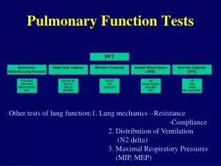
Routine PFT’s • Spirometry with or without Flow Volume loop • Static lung volumes • Single Breath Diffusing Capacity
Spirometry • Forced vital capacity • Forced Expiratory Volume in one second (FEV1) • Percent Expired (FEV1/FVC or FEV1%) • Forced Mid-Expiratory Flow (FEF 25-75) or Maximal Mid-Expiratory Flow (MMEF or MMF) • Peak or Maximal Expiratory Flow Rate (PEF or MEFR)
Obstructive Vent defect FVC reduced or Normal FEV1 reduced FEV1/FVC is reduced Example: Asthma, COPD Restrictive Vent defect FVC reduced FEV1 normal or reduced FEV1/FVC is increased Example: pulmonary fibrosis, pleural effusion, neuromuscular Pattern of defects seen on PFT’s
Lung Volumes • Functional Residual Capacity • Expiratory Reserve Volume • Residual Volume • Inspiratory Capacity • Total Lung Capacity • Vital Capacity
Diffusion • Transfer of a gas across a tissue sheet, governed by Fick’s law • Rate of Transfer = A D x P/T
Diffusion Capacity (measurement) A D x (P1- P2) T AD/T = Diffusion constante Rate of transfer (CO) = Vco = Dlco x (P1-P2) Dlco = Vco/ PA –Pa = Vco/ PA 25 ml/min/mmHg
Diffusing Capacity • Influenced by: • Changes in alveolar-capillary membrane • Pulmonary circulation • Ventilation perfusion matching • Hemoglobin concentration
Diffusion Capacity • Very important in • Interstitial lung disease • Drug induced lung injury • Reduced in Emphysema because of destruction of alveolar units
PFT Patterns in Disease PFT results are best interpreted with knowledge of the patients history, physical exam and occasionally chest X-ray.
Case 1 • 14 year old boy came to ER with increasing shortness of breath • History of asthma since age of 2-3 • Maintained on ICS and Beta2 agonists • Followed by Family physician, past year frequent attacks, several courses of antibiotics and systemic corticosteroids
Case 1 (cont’d) • In ER started on iv steroids and inhaled Beta 2 agonists, no improvement, admitted • No history of atopy, no nasal nor GI symptoms, no family history of asthma • Exam: decrease breath sounds • Admitted
Case 1 (cont’d) • CXR, CBC, chemistry non revealing • After 2 days of treatment with steroids and inhaled bronchodilators there was no improvement in symptoms • Noticed faint voice and tachypnea on minimal exercise • PFT obtained
Case PFT’s • FVC 93%, FEV1 45%, FEV1/FVC 41% • TLC 90%, RV 90%, DLCO 100% • ?????
Case 1(cont’d) • FOB: subglottic stenosis (? Congenital) • Tracheostomy followed by reconstructive surgery • Total recovery, no more asthma treatment
Case 2 • 32 year old man presented with 2 months history of increasing shortness of breath • Married, non-smoker, bank employee, no history of asthma • No other symptoms • Shortness of breath increasing before presentation • Seen by multiple physicians, given a number of antibiotics, bronchodilators, aminophylline
Case 2 (Cont’d) • Exam: BP 120/80, RR 18, P100, BMI 29, afebrile, chest: clear… rest of exam was normal • ER: ABG’s normal, CXR: normal, CT angio: normal, neuro consult (fellow): no neuro problem • Patient reassured by the team
Case 2 (cont’d) • Spirometry obtained: • FVC 50% • FEV1 55% • FEV1/FVC 80% • MVV 20% • ????
Case 2 (cont’d) • Neurology attending reconsulted • EMG: Myasthenia Gravis • Diagnosis suspected from FVC and MVV • Neuromuscular illness
Case 3 • A 60 year old man with history of ex-smoking, history of seasonal colds, admitted for hernia operation • Pulmonary consulted for pre-op clearance because of obesity • The patient denied pulmonary complaints, but his wife disclosed that he has a chronic cough
Case 3 Obstructed defect
Pre-operative screening • Patients with known pulmonary illness or symptoms • Overweight patients • Patients undergoing surgery in the chest or near the diaphragm
Case 4 • A 65 year old man non-smoker, lawyer, admitted for elective Lap Chole. Reports long history of mild cough, and dyspnea on exertion • Physical exam: bibasilar dry crackles (velcrow), clubbing of the fingers
Case 4 Restricted defect
Case 4 • TLC 60% • RV 40% • DLCO 40% • HRCT
Case 5 • 68 year old man with progressive dyspnea of one year duration, ex-smoker, no cough, no wheezing, no orthopnea… • History of CAD, SVT post angioplasty on multiple medication • EF% 55 • Meds: Plavix, beta one blocker, diuretics, cordarone, ARB,
Case 5 • FVC 50% • FEV1 55% • FEV1/FVC 85% • TLC 70% • DLCO 50%
Case 5 • PFT’s: Major drop in FVC and DLCO compared to the PFT done 2 years earlier • HRCT of chest: Increased markings over the bases, with areas of increased enhancement…. Consistent with Amiodarone toxicity
Follow up patients • Connective Tissue diseases (e.g. scleroderma) • Patients on Therapy that might affect the pulmonary system • Neuromuscular diseases