Download
1 / 1
10 likes | 189 Views
NYSBC MICRODIFFRACTION BEAMLINE (NYX). Group Leader : Wayne Hendrickson Proposal Team : R. Abramowitz 1 , W.A. Hendrickson 1,2,3 , J.P. Lidestri 1,2 , Q. Liu 1 , J. Schwanof 1 , X. Yang 1 1 New York Structural Biology Center, 2 Columbia University, 3 Brookhaven National Laboratory.

E N D
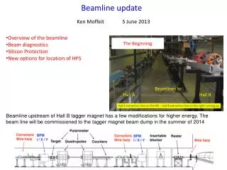
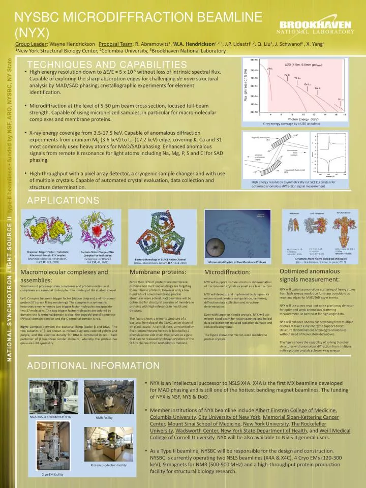
NYSBC MICRODIFFRACTION BEAMLINE (NYX) Group Leader: Wayne Hendrickson Proposal Team: R. Abramowitz1, W.A. Hendrickson1,2,3, J.P. Lidestri1,2, Q. Liu1, J. Schwanof1, X. Yang1 1New York Structural Biology Center, 2Columbia University, 3Brookhaven National Laboratory TECHNIQUES AND CAPABILITIES • High energy resolution down to ∆E/E = 5 x 10⁻⁵ without loss of intrinsic spectral flux. Capable of exploring the sharp absorption edges for challenging de novo structural analysis by MAD/SAD phasing; crystallographic experiments for element identification. • Microdiffraction at the level of 5-50 μm beam cross section, focused full-beam strength. Capable of using micron-sized samples, in particular for macromolecular complexes and membrane proteins. • X-ray energy coverage from 3.5-17.5 keV. Capable of anomalous diffraction experiments from uranium MV (3.6 keV) to LIII (17.2 keV) edge, covering K, Ca and 31 most commonly used heavy atoms for MAD/SAD phasing. Enhanced anomalous signals from remote K resonance for light atoms including Na, Mg, P, S and Cl for SAD phasing. • High-throughput with a pixel array detector, a cryogenic sample changer and with use of multiple crystals. Capable of automated crystal evaluation, data collection and structure determination. X-ray energy coverage by a U20 undulator High energy resolution asymmetrically cut Si(111) crystals for optimized anomalous diffraction signal measurement Applications TorT/TorS Sensor HK9 Sensor CysZ Transporter C2, 7-xtls, 2.3 Å 22 S / 492aa <ΔF>/<F> ~ 1.12% C2221, 13-xtls, 2.8 Å 28 S / 1162aa <ΔF>/<F>: ~ 0.83% I4122, 6-xtal, 2.3 Å 3 S / 157aa <ΔF>/<F> ~ 0.74% Chaperon Trigger Factor – Substrate Ribosomal Protein S7 Complex (Martinez-Hackert & Hendrickson, Cell 138, 923, 2009) Bacteria Slider Clamp – DNA Complex for Replication (Georgescu …O’Donnell Cell 132, 43, 2008) Structures from Native Biological Molecules (Liu … Hendrickson, Science, in press, 2012) Bacteria Homology of SLAC1 Anion Channel (Chen …Hendrickson, Nature 467, 1074, 2010) Micron-sized Crystals of Two Membrane Proteins Optimized anomalous signals measurement: NYX will optimize anomalous scattering of heavy atoms from high energy resolution for sharp transitions at resonant edges for MAD/SAD experiments. NYX will use a zero read-out noise pixel array detector for optimized weak anomalous scattering measurement, in particular for high angle data. NYX will enhance anomalous scattering from multiple crystals at lower x-ray energy to support direct structure determinations of biological molecules without need of heavy atom derivatives. The figure shows the capability of solving 3 protein structures with anomalous diffraction from multiple native protein crystals at lower x-ray energy. Macromolecular complexes and assemblies: Structures of protein-protein complexes and protein-nucleic acid complexes are essential to decipher the mystery of life at atomic level. Left: Complex between trigger factor (ribbon diagram) and ribosomal protein S7 (space filling rendering). The complex is a symmetric heterotetramer, whereby two trigger factor molecules encapsulate two S7 molecules. The two trigger factor molecules are colored by domain: the N-terminal domain is blue, the peptidyl-prolylisomerase (PPIase) domain is green and the C-terminal domain is red. Right: Complex between the bacterial clamp loader β and DNA. The two subunits of β are shown as ribbon diagrams colored yellow and purple, and the electron density for DNA is contoured in red. Each protomer of β has three similar domains, whereby the protein has quasi-six-fold symmetry. Microdiffraction: NYX will support routine structure determination of micron-sized crystals as small as a few microns. NYX will develop and implement techniques for micron-sized crystals manipulation, centering, diffraction data collection and structure determination. Even with larger or needle crystals, NYX will use micron-sized beam for raster scanning and helical data collection for reduced radiation damage and reduced background. The figure shows the micron-sized membrane protein crystals. Membrane proteins: More than 30% of proteins are membrane proteins and most known drugs are targeting to membrane proteins. However only a few hundreds of novel membrane protein structures were solved. NYX beamline will be optimized for structural analyses of membrane proteins with high relevance in health and diseases. The figure shows a trimeric structure of a bacterial homolog of the SLAC1 anion channel on plant leaves. A central pore, surrounded by five transmembrane helices, is blocked by a phenylalanine side chain that serves as a gate that can be released by phosphorylation of the SLAC1 channel from Arabadopsis thaliana. ADDITIONAL INFORMATION • NYX is an intellectual successor to NSLS X4A. X4A is the first MX beamline developed for MAD phasing and is still one of the hottest bending magnet beamlines. The funding of NYX is NSF, NYS & DoD. • Member institutions of NYX beamline include Albert Einstein College of Medicine, Columbia University, City University of New York, Memorial Sloan-Kettering Cancer Center, Mount Sinai School of Medicine, New York University, The Rockefeller University, Wadsworth Center, New York State Department of Health, and Weill Medical College of Cornell University. NYX will be also available to NSLS II general users. • As a Type II beamline, NYSBC will be responsible for the design and construction. NYSBC is currently operating two NSLS beamlines (X4A & X4C), 4 Cryo EMs (120-300 keV), 9 magnets for NMR (500-900 MHz) and a high-throughput protein production facility for structural biology research. NSLS X4A, a precedent of NYX NMR facility Protein production facility Cryo-EM facility