Download
1 / 17
170 likes | 447 Views
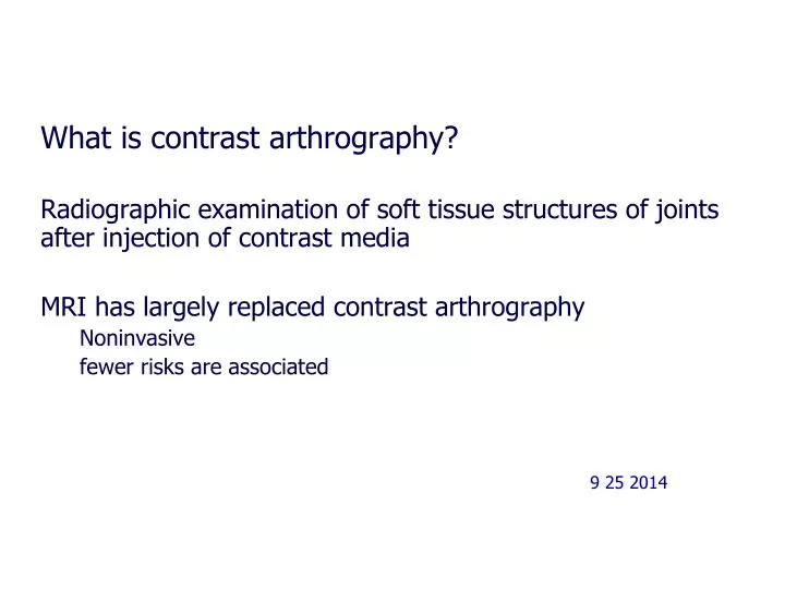
What is contrast arthrography? Radiographic examination of soft tissue structures of joints after injection of contrast media MRI has largely replaced contrast arthrography Noninvasive fewer risks are associated 9 25 2014. Contrast Media. Air Negative contrast Iodinated contrast

E N D
What is contrast arthrography? Radiographic examination of soft tissue structures of joints after injection of contrast media MRI has largely replaced contrast arthrography Noninvasive fewer risks are associated 9 25 2014
Contrast Media Air Negative contrast Iodinated contrast Positive contrast
Most common joints investigated using contrast arthrography • Knee • Hip • Shoulder • Wrist • TMJ
KneeProcedure Indications: • Cartilage, capsular injuries • Ligament or menisci injuries • Loose bodies • Joint rupture • Baker’s cyst • Synovial disease • Prosthesis check
Contraindications(Applies to all arthrograms) Skin infections Bleeding tendency Anti-coagulent therapy Allergy to contrast
Procedure Skin is first cleaned with betadine Local anesthetic is introduced Joint is punctured with needle (synovial fluid may be aspirated and sent for analysis)
Procedurecont’d Contrast introduced through needle under fluoroscopic guidance Needle is removed Pt asked to exercise knee to distribute contrast Pt is then turned prone if vertical method is used Spot radiographs are obtained by Radiologist Overheads
Vertical ray method Beam shoots down (or up if using fluoroscopy) Limb placed in stress device to widen side of jt space under investigation
Horizontal ray method To demonstrate lateral and medial meniscus Same procedure as with Vertical method but with crosstable CR (fluor not possible) Tear in medial meniscus Normal
Hip Arthrography Acetabular region of pelvis What kind of joint is hip? Diarthrotic- ball and socket joint Allows flexion, extension, abduction, adduction, rotation
Hip Arthrography cont’d Most often performed on children to evaluate hip dislocation before treatment In adults, primarily to check prosthesis dislocation or presence of infection Common puncture site: ¾ distal to inguinal crease and ¾ lateral to palpated femoral pulse
Radiographs AP Internal and external rotations Frog lateral
TMJ Arthrography CT and MRI have largely replaced Indications for Arthrogram of TMJ Diagnosing abnormalities of articular disk Subluxation (Incomplete or partial dislocation of bone in joint) Aplasia- (defective development) Fx. Ankylosis- (stiffness of jt) Arthritis
TMJ Arthrography Procedure: • Contrast injected ½ anterior to tragus of ear • Fluoroscopy used to observe and image mandibular motion • Radiographs made with pt’s mouth closed, partially open, and fully open
Shoulder Arthrography Indications: • Rotator cuff or long head of biceps tears • Foreign bodies • Persistent pain or weakness • Frozen shoulder After injection, take standard shoulder projections CT often used in conjunction with shoulder arthrograms
Shoulder Arthrograpy Normal Rotator Cuff Tear
Wrist Arthrography Indications Trauma Persistent pain Limitation of motion Procedure Contrast injected into dorsal wrist Wrist manipulated to disperse contrast Routine images