Download
1 / 20
230 likes | 535 Views
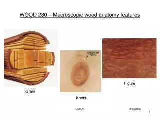
WOOD 280 Wood Anatomy and Identification Dr. Simon Ellis. Softwoods. Hardwoods. Lodgepole pine. Aspen. Oak. Hemlock. Douglas-fir. Spruce. Birch. Maple. (Waddington arboretum). May 21. May 3. October 11. December 20. (Ellis). Tree trunk showing the successive concentric layers.

E N D
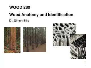
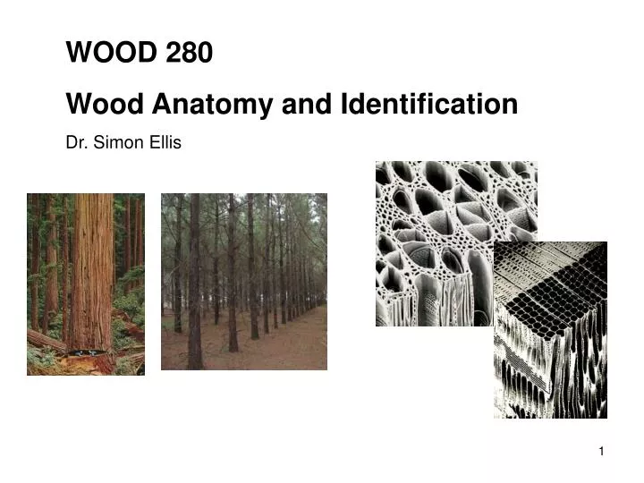
WOOD 280 Wood Anatomy and Identification Dr. Simon Ellis
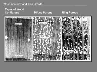
Softwoods Hardwoods Lodgepole pine Aspen Oak Hemlock Douglas-fir Spruce Birch Maple (Waddington arboretum)
May 21 May 3 October 11 December 20 (Ellis)
Tree trunk showing the successive concentric layers Outer bark - dead tissue that protects the inner tissues from drying out, from mechanical injury and from insects Inner bark (phloem) – conducts sugars produced by photosynthesis to the roots and other non-synthetic parts of the tree Cambium – produces secondary xylem and secondary phloem Sapwood – consists of xylem tissues through which water and minerals move from the soil to the leaves and other living parts of the tree Heartwood – composed entirely of dead cells, supporting column of the mature tree (St. Regis Paper Company)
Sapwood - Heartwood Sapwood Heartwood (Hoadley) (Core, Côté & Day)
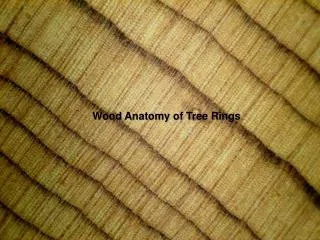
earlywood latewood (Hoadley)
Three-dimensional representation of the vascular cambium (Haygreen and Bowyer)
Cambial cell division (Haygreen and Bowyer)
Schematic drawing portraying ontogeny of young tree stem c cortex d epidermis e epidermis pc procambium p pith pp primary phloem px primary xylem vc vascular cambium sp secondary phloem sx secondary xylem (Panshin and de Zeeuw)
Cell development at apical shoot Epidermis Protoderm Primary phloem Secondary phloem Apical initials Mother cells Procambium Vascular cambium Primary xylem Secondary xylem Cortex Ground meristem Pith
Representation of developing stem (Haygreen and Bowyer)
Portion of a transverse section of a young stem showing arrangement of tissues • Mature xylem • Zone of xylem differentiation • Cambial zone • Zone of phloem differentiation • Mature phloem 1 2 3 4 5 (Zimmerman and Brown)
Periclinal division of cambial fusiform initials (Haygreen and Bowyer)
Anticlinal division of cambial fusiform initials (Panshin and de Zeeuw)
Formation of new ray initials in the vascular cambium (a) (b) (c) (d) (e) (f) • (a) Initial awith extensive ray contact survives, while initial b with sparse ray contact matures into a deformed cell and disappears • A ray is split by instrusive growth of a fusiform initial • A new ray initial arising from pinching off the top of a fusiform initial • Two single ray cells are formed through reduction of a short fusiform initial; either or both of these cells may survive and later develop into rays consisting of a number of cells formed by subsequent division of these initials or they may be eliminated • A new ray is formed by septation of the entire short fusiform initial • A new ray initial is formed on the side of a fusiform initial, which will continue to function as such (Panshin and de Zeeuw)
Plant Hormones – nature, occurrence and effects (Raven, Evert & Eichorn)