Download
1 / 46
460 likes | 473 Views
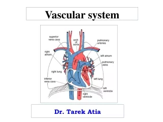
Cardio-vascular system diseases. Associate professor Golovata T. Cardiovascular disease is a class of diseases that involve the heart or blood vessels (arteries, capillaries and veins) and is leading cause of death in Europe and North America.

E N D
Cardio-vascular system diseases Associate professor Golovata T.
Cardiovascular disease is a class of diseases that involve the heart or blood vessels (arteries, capillaries and veins) and is leading cause of death in Europe and North America. In the UK in 2009, around one third of all deaths in the UK were due to cardiovascular disease. Of these, over 82,000 deaths were caused by coronary heart disease, and about 49,000 were caused by stroke.
Types Cardiovascular disease includes: • coronary heart disease (heart attacks) • cerebrovascular disease (stroke) • raised blood pressure (hypertension) • peripheral artery disease • rheumatic heart disease • congenital heart disease and heart failure • valvular heart disease • cardiomyopathy - diseases of cardiac muscle • inflammatory heart disease
Risk factors • Almost all cardiovascular disease in a population can be explained in terms of a limited number of risk factors: age, gender, high blood pressure, high serum cholesterol levels, tobacco smoking, excessive alcohol consumption, family history, obesity, lack of physical activity, psychosocial factors, diabetes mellitus, air pollution.
Epidemiology The most studied mortality from cardiovascular diseases as a manifestation of generalized atherosclerosis. In the Russian Federation in 2000, deaths from cardiovascular diseases was 800.9 per 100 000 population. For comparison: France - 182.8 (the lowest in Europe); Japan - 187.4. It is shown that reducing the risk of cardiovascular disease in these countries is concerned not so much with the quality of medical care as a way of life and eating habits.
According to the WHO definition atherosclerosis - a "diverse mix changes the inner lining of the arteries, which manifest as focal deposits of lipids, complex carbohydrates compounds, and elements of blood circulating in the matter, the formation of connective tissue and calcium adjournment." Atheroma (Atherosclerosis)
Deposits formed atheromatous plaques. The next expansion in their connective tissue (sclerosis) and calcification of the vessel wall leading to deformity and lumen narrowing up to their obliteration (obstruction).
Etiology. Causes of atheroma is not known. There are several theories of atheroma • Theory lipoprotein infiltration - initial accumulation of lipoproteins in the vascular wall; • Theory of endothelial dysfunction - primary violation of protective properties of the endothelium and its mediators; • Autoimmune - primary dysfunction of macrophages and leucocytes infiltration of their vascular wall; • Monoclonal - initial clone of abnormal smooth muscle cells;
Pathogenesis of atherosclerosis Pathogenetic essence of atherosclerosis is the focal putting off in the intima of the arteries so-called atherogenic lipoproteins. Lipoproteins are spherical particles consisting of a core and outer shell. In the nucleus consists of triglycerides and cholesterol esters, the composition of the outer shell - proteins, phospholipids and cholesterol.
Pathogenesis of atherosclerosis Atherosclerosis is a multifactorial disease that usually develops many years before any clinical symptoms are manifest. Clinical events include ischemic heart disease (coronary arteries), arterial occlusive disease (peripheral arteries), stroke (cerebral arteries), kidney failure (renal arteries), and aortic aneurism (aorta)
Morphogenesis of atherosclerosis Macroscopically distinguish the following stages: Yellow spots or bands; fibrous plaque Stage complicated changes (ulceration, calcification, thrombosis)
The first stage of atherosclerosis is endothelial damage and dysfunction, which stimulates the accumulation and oxidation of LDL-C in the vessel wall. Monocytes migrate from the blood into the subendothelial intima and transform into macrophages, which accumulate lipids (foam cells) to form the lipid core of the atherosclerotic plaque. Morphogenesis of atherosclerosis Microscopic manifestations
Production of inflammatory mediators and cytokines stimulate migration and proliferation of smooth muscle cells of the vascular intima, and deposition of extracellular matrix molecules such as elastin and collagen, which leads to plaque expansion and the formation of the fibrous cap. Morphogenesis of atherosclerosis Liposclerosis
The cholesterol clefts of lipid, along with a few scattered foam cells and a couple of lymphocytes, are seen at high magnification in this atheromatous plaque. Morphogenesis of atherosclerosis
Atheromatous plaques may undergo a series of changes resulting in complicated plaques. These include calcification, ulceration, overlying thrombus formation, haemorrhage (as seen in this case) and aneurysmal dilatation of the vessel. It is these changes that usually account for the serious clinical consequences of this disease. Morphogenesis of atherosclerosis
Atherosclerosis of the aorta-macroscopic pathology • It shows multiple variable sized atheromatous plaques which become confluent in the abdominal aorta. These plaques are well circumscribed, slightly raised and yellow/white in colour. Some of the larger plaques are complicated by superficial ulceration with adherent thrombus and focal dystrophic calcification.
Atherosclerosis is the most frequent cause of aortic aneurysms. Aneurysms is a focal stretching or ballooning of the aorta. Usually occur in the abdominal aorta When they reach a diameter of over five centimeters (about two inches), the risk of fatal rupture increases. A rupturing aneurysm may cause symptoms of abdominal pain or rigidity, rapid heart rate, nausea and anxiety. A rapid loss of blood pressure (shock) may follow. Sudden rupture is associated with a high death rate. Complication Rupture of aortic aneurysm
Defined as cerebrovascular disease. Manifestations are ischemic (usually) or hemorrhagic stroke Atherosclerosis of cerebral arteries
Blood flow to the extremities may be reduced because of this narrowing, and may not adequately provide for the need of oxygen for the tissues. Many patients experience leg pain, referred to as "intermittent claudication". complete occlusion causes gangrene Atherosclerosis of lower extremities
Atherosclerosis of the renal arteries Atrophy secondary to renal artery atherosclerosis: Gross, natural color, both kidneys one very atrophic and wrinkled. The large left kidney weighed 220 grams and the small left one 90 gram
HYPERTENSION (High Blood Pressure) Hypertension is the commonest cause of cardiac failure in many societies and a major risk factor for atherosclerosis. Furthermore, it is a major risk factor for cerebral haemorrhage, another leading cause of death worldwide. There is no universally agreed definition of hypertension, but most authorities would accept that a sustained resting blood pressure of more than 160/95 mmHg is definite hypertension
Aetiological classification • Essential (primary) hypertension: Genetic susceptibility. Excessive sympathetic nervous system activity. Abnormalities of Na/K membrane transport. High salt intake. Abnormalities in renin-angiotensin-aldosterone system.
Aetiological classification • Secondary hypertension: Chronic renal failure. Renal artery stenosis. Glomerulonephritis. Endocrine causes. Adrenal tumours (cortical or medullary). Cushing's syndrome. Coarctation of aorta.
Pathological classification Hypertension is classified also according to the clinicopathological consequences of the blood pressure elevation. Benign or essential hypertension is often asymptomatic and discovered only during a routine medical examination. Malignant hypertension is a serious condition necessitating prompt treatment to minimise organ damage or the risk of sudden death from cerebral haemorrhage
Vascular lesions in hypertension Aorta-elastofibrosis Arteries-hyperelastosis.Arteriolar- hyalinosis
The characteristic histological lesion of malignant hypertension is fibrinoid necrosis of small arteries and arterioles Hypertension
Changes in kidneys -Primary-wrinkled kidney • The underlying sclerosis and hyalinosis of arterioles renal glomeruli (1.2). Kidney wrinkled, its fine-grained surface.
CORONARY (ISCHEMIC) HEART DISEASE Ischemic heart disease (IHD), or myocardial ischaemia, is a disease characterized by ischaemia (reduced blood supply) of the heart muscle, usually due to coronary artery disease (atherosclerosis of the coronary arteries).
Classification of IHD Sudden coronary death. Angina Myocardial infarction Heart failure
The immediate cause of myocardial infarction • progressive atherosclerotic stenosis • erosion of an atheromatous plaque with superimposed thrombosis • rupture of the fibrous cap of a plaque with haemorrhage into the lesion and thrombosis • prolonged coronary spasm against her atherosclerotic lesions
This is thrombosis in a coronary artery. Such a thrombus severely narrows or occludes the lumen and can produce a sudden ischemic event. "Sudden death" as well as infarction can occur. Cause of myocardial infarction.Thrombosis of coronary artery, gross.
This severely narrowed coronary artery has the remaining lumen filled by thrombus. Cause of myocardial infarction.Thrombosis of coronary artery, microscopic.
Transmural: associated with atherosclerosis involving a major coronary artery. It can be subclassified into anterior, posterior, inferior, lateral or septal. Classification myocardial infarction
Subendocardial: involving a small area in the subendocardial wall of the left ventricle, ventricular septum, or papillary muscles. Classification myocardial infarction
Myocardial infarction. Morphology • 24 - 72 hours from onset. Total loss of nuclei and striations along with heavy neutrophilic infiltrate
Complications • Arrhythmias • Extension of infarction, or re-infarction • Congestive heart failure (pulmonary edema) • Cardiogenic shock • Pericarditis • Mural thrombosis, with possible embolization • Myocardial wall rupture, with possible tamponade • Papillary muscle rupture, with possible valvular insufficiency • Ventricular aneurysm formation
Complications • When the infarction is 3 to 5 days old, the necrosis and inflammation are most extensive, and the myocardium is the softest, so that transmural infarctions may be complicated by rupture. A papillary muscle may rupture as well to produce sudden valvular insufficiency. Rupture through the septum results in a left-to-right shunt and right heart failure.
A cross section through the heart reveals a ventricular aneurysm with a very thin wall at the arrow. Note how the aneurysm bulges out. The stasis in this aneurysm allows mural thrombus, which is present here, to form within the aneurysm. Complications
Cardiomyopatias • Cardiomyopathies are diseases of heart muscl. Cardiomyopathies include a variety of myocardial disorders that manifest with various structural and functional phenotypes and are frequently genetic. Although some have defined cardiomyopathy to include myocardial disease caused by known cardiovascular causes (such as hypertension, ischemic heart disease, or valvular disease), current major society definitions of cardiomyopathy exclude heart disease secondary to such cardiovascular disorders.
Cardiomyopatias-classification • Dilated cardiomyopathy (DCM) • Hypertrophic cardiomyopathy (HCM) • Restrictive cardiomyopathy (RCM) • Arrhythmogenic right ventricular cardiomyopathy/dysplasia (ARVC/D) • Unclassified cardiomyopathies
Illustration of dilated cardiomyopathy (right), showing a dilated left atrium and left ventricle, bulging interventricular septum from left to right, and thin ventricular walls. For comparison, a normal heart is shown on the left. Dilated cardiomyopathy
Illustrations of a normal heart (left) and a heart with hypertrophic cardiomyopathy (HCM). Note that the heart walls (muscle) are much thicker (hypertrophied) in the HCM heart. Hypertrophic cardiomyopathy
Illustration of dilated cardiomyopathy - fibrosis under the endocardium and in the the inner third of the myocardium. Restrictive cardiomyopathy

![CARDIO-VASCULAR SYSTEM [CVS] FUNCTIONAL ANATOMY OF HEART](https://cdn1.slideserve.com/1739818/cardio-vascular-system-cvs-functional-anatomy-of-heart-dt.jpg)