Download
1 / 23
240 likes | 268 Views
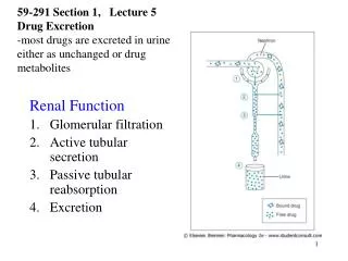
Glomerular Filtration Rate. Use of GFR to classify renal impairment 1. Decreased Renal reserve. When 50% of the nephrons are destroyed (One kidney). GFR drops to 50%. Homeostasis is perfectly maintained. Urea and creatinine can be within the normal range.

E N D
Use of GFR to classify renal impairment • 1. Decreased Renal reserve. When 50% of the nephrons are destroyed (One kidney). GFR drops to 50%. Homeostasis is perfectly maintained. Urea and creatinine can be within the normal range. • 2. Renal Insufficiency: When GFR drops to 20-50%. The earliest sings is isosthenuria or polyuria with isotonic urine. Azotemia, anemia, and hypertension appear too. • 3. Renal Failure: GFR drops to less than 20% N. All signs and symptoms of uremia (urine in the blood) are present. • 4. End-stage Renal Disease ESRD: Occurs when GFR drops to less than 5% N. At this stage, dialysis or transplantation are necessary for survival. Is an administrative term rather than medical term. It means that person should be covered by government insurance, because replacement therapy is mandatory.
GFR = [(140-age in yr) X (weight in kg)]/(72 X serum creatinine in mg/dl). • Values for women are 85% of the predicted. • 186 (serum creatinine in mg/dL)-1.154 (age in years)-0.203 • Schwartz Formula in Children • GFR (mL/min/1.73 m2) = k * Height (cm) / Serum Creatinine (mg/dl) • k = Constant. • k = 0.33 in premature Infants • k = 0.45 in Term infants to 1 year old • k = 0.55 in Children to 13 years • k = 0.65 in Adolescent males….in females it remains 0.55
Estimation of GFR in a pediatric patients • The Schwartz Equation • GFR can be estimated in a pediatric patient simply using plasma creatinine without all the hassle of 24 hour urine collection. • The equation is eGFR = (k * height) / Pcr (in mg/dl)
Estimation of GFR in a pediatric patients Example: for 6-y old child: k = 0.55, h = 110cm, Pcr = 0.33mg/dl • eGFR = (0.55 * 110) / 0.33 = 183ml/min/1.73m2
Estimating GFR in adults • Let us take the example of an 85 year old geriatric female patient. • Weight = 75kg, Pcr = 1.5mg/dl • eGFR = [(140 – 85) * (75kg)] * 0.85 / (1.5 * 72) = 36ml/min/1.73 m2
Estimating GFR in adults • The bottom line is that equations for estimation of GFR are available and accurate. Pcr and anthropometric measures are utilized without the need for 24 hour urine collection. • Gradual loss of renal function with age is a normal process (1% each year), as in the case of the female patient in the previous example. Although her GFR is markedly reduced, it is probable that she has normal renal function. Even if she has hypertension, it is most likely due to age-related vascular degenerative processes. • Notice that estimations of GFR are unacceptable in cases of end-stage renal disease.
Implications of measuring GFR • Many chronic diseases affect renal function. One major example is diabetes mellitus, which causes micro-angiopathy in the renal vasculature. This in turn alters glomerular filtration. • You might have noticed from the example above that the GFR is way below normal, even though Pcr is still at the upper limit of normal range. The reason is that serum creatinine remains around normal even when the GFR is reduced to less than half of its normal value. Kidney impairment is present, but it is not serious.
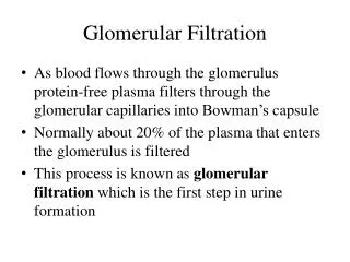
Filtration in systemic capillary beds VS Glomerular Filtration • filtration across the systemic capillary bed of capillaries is only 20L/day ;17L is reabsorbed by veins and 3L by lymphatics (kidneys are not included). • filtration through the glomerular capillaries is 180L/day;i.e., 9 times more than the systemic filtration. Why?
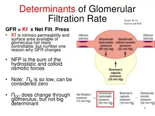
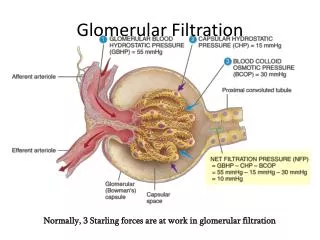
GFR=Kf.[(PGC-PBS)-(GC- BS)]=Kf * Peff (use Ohm’s law again) • The driving force is the summation of Starling forces which are 2 forces inside and only one force outside. • The inside ones are: • Capillary hydrostatic pressure (PGC). • Colloid capillary pressure (GC) provided by albumin and globulin mostly by albumin. • The outside ones are: • PBS; it is 18 mmHg (encapsulated organs) and it will oppose filtration ( in most tissue interstitial pressure is subatmospheric or –ve)
Dirving Forces affecting Filtration • Favoring Filtration: • Hydrostatic Pressure in the Glomerular capillaries. (PGC) =60 mmHg..the highest in our body • Oncotic (Colloid) Pressure of the filtrate in the Bowman’s capsule. (BS)=32 mmHg • Opposing Filtration: • Hydrostatic Pressure in the Bowman’s capsule.(PBS)=18 mmHg
I.Glomerular Hydrostatic Pressure: • The difference between 20L/day (systemic capillaries) and the 180L/day (GFR) is either due to increased Peff or increased Kf or both. • The PGChere is 60 mmHg as opposed to 15-30 mmHg in systemic capill, or 7-10 mmHg in pulmonary capillaries…..WHY? • If we look at systemic capillaries they have an arterial end and a venous end but in the glomerulus they have both arteriolar ends afferent and efferent arterioles. This makes the pressure in the glomerular capillaries the highest (60 mmHg).
Arteriolar diameter effect on GFR: • Afferent dilatation means an increase in the blood coming to the capillaries so increased PGC and GFR. • Constriction of efferent arteriole increases PGC to a limit. If it goes over this limit filtration will decrease as no more blood entering the capillaries. • To regulate PGC you either control the afferent arteriolar dilatation or the efferent arteriolar constriction or both.
II. Glomerular Capillaries Oncotic(colloid) Pressure: • In the systemic capillaries the stays 28mm Hg at both the arterial and venous ends. WHY? Because what is filtered is only 0.5% from the whole incoming fluid, so it does not affect the concentration of proteins much. • But filtration in the kidneys is 20% so it must have an effect so GC that becomes 36mm Hg at the efferent end (avg. 32 mmHg).
Interstitial forces (Bowman’s Space) : • Bowman’s Space contains protein free glomerular filtrate; i.e, too small BS. So in the kidneys Starling forces have been reduced to 3 forces instead of 4 forces • Hydrostatic Pressure (P) of Bowman’s space is 18 mmHg due to the fluids filtered. • Net Driving forces favoring filtration= 60 – (32 + 18 ) = 10 mmHg • Knowing that P = 10mmHg and GFR is 125 then Kf will equal =12.5 ml/min.mmHg • (125=10*KF)
Renal Kf Basement membrane is negatively charged • In nephrotic syndrome, loss of negative charge causes albumin loss and edema. • (Remember the four cause of hypoalbuminemia: malnutrition, malabsorption, malproduction, and increased loss from the kidney). • Hypoalbuminemia →↑GFR.
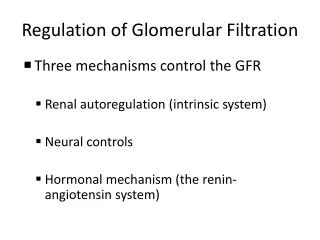
Renal Autoregulation • Autoregulationof GFR…see the figure after 3 slides • GFR is fluctuates slightly in relation to changes in arterial blood pressure but this translates in a large increase in urine output… why is that? • GFR = 125ml/min and UOP is only = 1ml/min = 1.5L/day which means 124ml/min is reabsorbed (99.4% of the filtered water is reabsorbed and only 0.6% is excreted) so a little change in GFR changed the urine output a lot. • Therefore, GFR must be regulated and this is achieved mainly by the renal vascular system (glomerular capillary hydrostatic pressure) and this is controlled by afferent and efferent arterioles by the following mechanism:
Tubuloglomerular Feedback • In the distal tubule the afferent arteriole touches a few cells in its wall…these DT cells are sensors (macula densa). They sense the content of Na+, K+, Ca++ . When the amount of these electrolytes reach macula densa is small → 2 messages are sent. • The first message: dilatation of the afferent arteriole so the blood flow increases to the capillaries. (Myogenic Response) • The second message: is to the granular cell in the afferent arteriole to secret rennin (hormonal response). • Rennin leaves the kidney and goes to the circulation where it cleaves 4 aminoacids for a 14-aa small peptids produced by the liver and called angiotensinogen forming the decapeptide angiotensin-I (decapeptide). In the lungs the ACEs (angiotensin converting enzymes) conver AI to AII (octapeptide). • In bleeding we have to protect the kidneys by keeping normal GFRand at the same time conserve water. Angiotensin-II can do this…through:
Tubuloglomerular Feedback • First: constriction of efferent arteriole leading to increased GFR and at the same time the pressure in the peritubular capillaries decreases giving a better chance for reabsorbing to get the minimal urine output which is 0.5L/day. • Second: angiotensin acts directly on the adrenal cortex to secret aldosterone that enhances the reabsorption of Na+ from the distal tubule and sodium bring with it water. • Third: angiotensin itself act directly to enhance sodium reabsorption in the proximal tubule.