Download
1 / 48
480 likes | 622 Views
NUTRITION. How Organisms Obtain Nutrition. AUTOTROPHS Examples are green plants, algae, some microorganisms Most are photosynthetic Using sunlight , CO 2 , water food Are called phototrophs Some bacteria are chemosynthetic Using energy from chemical reactions to produce food

E N D
How Organisms Obtain Nutrition AUTOTROPHS Examples are green plants, algae, some microorganisms • Most are photosynthetic • Using sunlight, CO2, water food • Are called phototrophs • Some bacteria are chemosynthetic • Using energy from chemical reactions to produce food • Are called chemotrophs
How Organisms Obtain Nutrition HETEROTROPHS • All animals • Some microorganisms
Energy content of food • Energy is provided to the organisms by the chemical breakdown of carbo’s, fats, and proteins • Energy is released and stored as ATP in certain molecules during the process of cellular respiration • The measure of this energy released by cellular respiration is in calories • Energy released by 1 gram of a carbohydrate = 4 kilocalories • Energy released by 1 gram of a protein = 4 kilocalories • Energy released by 1 gram of a fat = 9 kilocalories
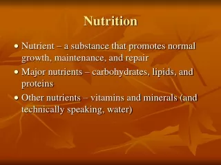
Humans need 6 nutrients • Proteins • Carbohydrates • Fats • Vitamins • Minerals • Water
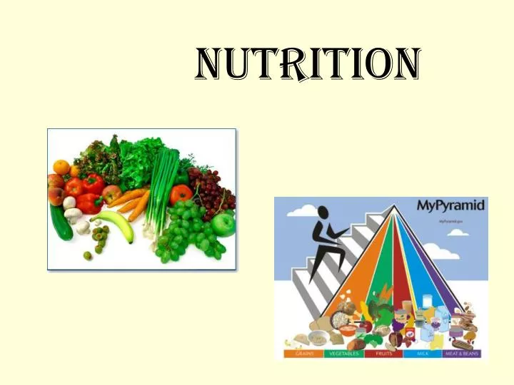
Four (five) basic food groups • Milk and cheese • Meat, poultry, fish, beans Legumes – plants that have seeds w/ pods Beans, peas, peanuts are high in proteins • Fruit, vegetables • Bread and cereals • Sweets • Fiber • bulky indigestible materials including cellulose from the cells walls of fruits, vegetables, and grains • fiber stimulates the digestive muscles • decreases the risk of colon cancer
Mechanical breakdown vs. chemical breakdown of food • Mechanical breakdown = foods are broken, crushed into smaller pieces with out being changed chemically. This gives the materials a greater surface area so that the digestive enzymes can work more efficiently on them.
Mechanical breakdown vs chemical breakdown of food • Chemical breakdown (digestion) = carried out by digestive enzymes breaking down large molecules into smaller and smaller ones by chemical reactions. • Example: hydrolysis
Nutrition in an Ameba • Has pseudopods “false feet” • Surrounds food when it contact with it • The cell membrane forms a food vacuole • Food vacuole joins with a lysosome and digestive juices break down the food particle • Small particles diffuse out of the vacuole into the cytoplasm • Indigestible materials remain in the vacuole until the contents are expelled from the cell
Ameba ingestion http://www.youtube.com/watch?v=W6rnhiMxtKU
Nutrition in a paramecium • Cilia sweep food particles down the oral groove • Into the gullet which pinches off to form a form a food vacuole • Food vacuole joins with a lysosome and digestive juices break down the food particle • Usable materials diffuse into the cytoplasm • Indigestible materials are discharged from the cell through the anal pore
Nutrition in a paramecium http://www.youtube.com/watch?v=saLYHUs6cWk
Nutrition in a hydra • Performs intra- and extracellular digestion • Threads in the tentacles wrap and poison prey • Captured food is pushed into the oral groove • Endoderm cells secrete digestive enzymes into the gastrovascular cavity (extracellular digestion) • Nutrients are absorbed into the cells forming food vacuoles (intracellular digestion) • Wastes can diffuse two ways: • Out the ectoderm into the outside environment • Out the endoderm into the gastrovascular cavity and out the mouth
Nutrition in a hydra http://www.youtube.com/watch?v=zkF_1r6ll54&feature=related
Nutrition in an earthworm • Digestive tube called an alimentary canal • Food is sucked into the mouth by a muscular pharynx to • Esophagus to • Crop for storage to • Gizzard for grinding (mechanical digestion) • To the intestine for chemical digestion and absorption • Indigestible material pass out through the anus
Nutrition in an earthworm • A typhlosole is a fold in the digestive wall • It increases surface area • Therefore increases absorption
Nutrition in a grasshopper • Mechanical digestion by mouth parts • Salivary glands (saliva) secrete digestive enzymes • To the esophagus to the crop (storage) • To the gizzard (mechanical digestion) • To the stomach (chemical digestion and absorption) • Indigestible (dry) materials go through the intestine out the anus • Water is absorbed in the rectum
Grasshopper eating a leaf http://www.youtube.com/watch?v=F3JJFSsVUBg
Contact and surround food • Membranes join • Food vacuole forms • Vacuole fuses with lysosome Pseudopod (false feet) • Cilia sweep food into oral groove • Gullet pinches off into vacuole • Vacuole fuses with lysosome Cilia • Poisons prey • Tentacles capture food and push it into the mouth • Exrtacellular digestion in gut • Absorption into the endoderm Special stinging cells Wrapping Treads out of tentacles Alimentary canal Typhlosole (↑ surface area) Mouth – pharynx-esophagus-crop (storage)–gizzard (mech dig) – intestine (chem dig and absorption) Mouth parts (mech dig) –salivary glands (chem dig) – esophagus – crop (storage) – gizzard (mech dig) – stomach (chem dig and absorption) – intestine – rectum (water absorption) – anus (dry waste) Tube in a tube digestive system
The Human Digestive System • Complete the diagram on page 14 in your note packet
The Human Digestive System • Mouth • Tongue • Salivary glands • Epiglottis • Esophagus • Liver • Stomach • Pancreas • Large intestine • Small intestine • Appendix • Rectum • Anus
Accessory organs • Stomach (not seen here) • Pancreas • Gallbladder • Liver • (Common) Bile duct • Duodenum • Pancreatic duct
The mouth and pharynx • teeth - mechanical digestion • tongue - moves and shapes the food mass • 3 pair of salivary glands secrete saliva • thin saliva wets food • thick saliva sticks food together into a bolus • contains digestive enzymes for chemical digestion • salivary amylase breaks down starch to maltose (a polysaccharide to a disaccharide) • tongue pushes food into the pharynx - starts swallowing reflex
The esophagus • flap of tissue called epiglottis prevents food from going down the trachea instead of the esophagus • peristalsis - alternating relaxation and contraction of the muscular walls • sphincter (ring of muscle) where the esophagus opens to the stomach called the cardiac sphincter
The stomach • mechanical digestion by contractions of stomach walls • chemical digestion by glands in stomach lining • pyloric glands - mucus to protect the stomach lining • gastric glands - acidic gastric juices • kills bacteria • contains pepsin to break down large proteins into polypeptides • solids form soupy liquid called chyme • pyloric sphincter opens to the small intestine
The small intestine • chemical digestion • pancreatic juice from the pancreas • bile from the liver • intestinal juice from glands in the walls • absorption • simple sugars, amino acids, vitamins, minerals absorbed through villi into the blood stream • increased surface area • very long • many folds • villi and microvilli • fatty acids and glycerol absorbed into lymphatic system
Intestinal villi • Intestinal villi (singular: villus) are tiny, finger-like projections that come out from the wall of the small intestine and have additional extensions called microvilli (singular: microvillus) which protrude from epithelial cells lining villi. They increase the absorptive area and the surface area of the intestinal wall. It is important that the food is absorbed at a considerably fast rate so as to allow more food to be absorbed. (If the process is too slow, the concentration of the blood in the blood vessels and the food will be equal, thus, diffusion will not occur.) Digested nutrients (including sugars and amino acids) pass into the villi through diffusion. Circulating blood then carries these nutrients away.
Intestinal villi Blood capillaries absorb amino acids, glucose, vitamins and minerals into the blood The lacteals absorb fatty acids and glycerol into the lymphatic system
peristalic movement • squeeze chyme through intestine • mix chyme with digestive juices • breaks down mechanically • speed absorption by pushing chyme against walls
pancreas • hormones from the stomach stimulate pancreas to secrete pancreatic juices and enzymes into the small intestine (duodenum) • pancreatic juice neutralizes stomach acid • pancreatic enzymes • amylase breaks down starch to maltose • proteases break down proteins • lipase breaks down fats
Gall bladder • stores bile made in the liver • bile travels from liver to gallbladder (storage) into bile duct to duodenum (small intestine) • bile emulsifies fat - bile has no enzymes
Large intestine • small pouch where large and small intestine join is the appendix • undigested and unabsorbed materials pass through large intestine • reabsorption of water from the food mass • absorbs vitamins produced by bacteria here • elimination - removal of undigested material (feces) • feces stored in the rectum and then defecated out the anus
Digestive glands • glands that secrete substances that aid in digestion • the food mass never passes through these glands
Digestive glands:the liver • produces bile • bile emulsifies fat • stores excess carbohydrates in the form of glycogen • bile is slightly basic and assists in neutralizing acid chyme from the stomach • bile stored in the GALLBLADDER • bile travels: GALLBLADDER BILE DUCT DUODENUM (small intestine)
Digestive glands:the pancreas • produces pancreatic juices • aids in digestion of • proteins • starch • fats • pancreatic juices are basic and neutralize the acid chyme from the stomach The small intestine is slightly basic (pH ~ 8)
Human nutrition page 20 in your packet
Begins chemical digestion of starch • Wets food • Sticks food together Salivary glands esophagus Transports food to the stomach stomach • Begins chemical digestion of protein • Mechanical digestion liver • Produces bile • Stores extra starch as glycogen Produces pancreatic juice that aids in chemical digestion of all food groups pancreas Gall bladder Stores bile Small intestine • Completes chemical digestion • Absorption of nutrients • reAbsorption of water • Production and absorption of vitamins Large intestine rectum Storage of feces
Which organs have a “S” on them? • Salivary glands • Stomach • Liver • Pancreas • Small intestine
Which organs have a “ D” on them? • Mouth (not shown) • Stomach • Small intestine
Salivary glands starch glands in stomach walls proteins emulsification of fats liver fats, starch, proteins pancreas glands in the intestinal wall fats, starches proteins
simple sugars or monosaccharides • The end products of the digestion of carbohydrates are • The end products of the digestion of proteins are • The end products of the digestion of lipids are amino acids 3 fatty acids and 1 glycerol
salivary glands salivary amylase starch or polysaccharides maltose disaccharide small amino acid chains activates pepsin stomach pepsin proteins Emulsifies fats in to droplets liver none fats trypsin → amylase → lipase → proteins → starch → fats → peptides maltose f. a. + glycerol pancreas glands in the intestinal wall peptidase → lactase → maltase → sucrase → peptides → lactose → maltose → sucrose → amino acids glucose glucose glucose