Download
1 / 1
10 likes | 88 Views
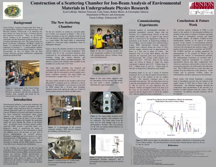
Construction of a Scattering Chamber for Ion-Beam Analysis of Environmental Materials in Undergraduate Physics Research Scott LaBrake, Michael Vineyard, Colin Turley, Robert Moore, & Christopher Johnson Department of Physics and Astronomy Union College, Schenectady, NY.

E N D
Construction of a Scattering Chamber for Ion-Beam Analysis of Environmental Materials in Undergraduate Physics Research Scott LaBrake, Michael Vineyard, Colin Turley, Robert Moore, & Christopher Johnson Department of Physics and Astronomy Union College, Schenectady, NY Conclusions & Future Work The ion-beam analysis technique of PIXE is well suited to the analysis of environmental pollution for elements in the range of sodium to uranium, especially those found in atmospheric aerosols and liquid precipitation samples. In addition, UCIBAL involves undergraduate students in all aspects of research projects on the analysis of environmental materials. An undergraduate team of researchers performed data collection and subsequent data analysis using a 1.0MV tandem electrostatic particle accelerator and GUPIX13. The UCIBAL provides an exciting experience for undergraduates to do research in Physics and in particular, the analysis of environmental materials. The undergraduate researchers are provided with a first-hand, hands-on introduction to research in experimental physics that fully complements their classroom experiences and provides a basis for an on-going project that they can really tackle and make their own. The UCIBAL provides an avenue that takes our students from the classroom to the laboratory. Union College and in particular UCIBAL values our undergraduates and their experiences in the laboratory. Future studies will include beginning a systematic study of atmospheric aerosols near two airports in the capital region of NYS and at Piseco Lake in the Adirondacks. An elemental mapping, showing the composition and concentrations of the aerosols as a function of radial distance from the source, will be constructed. In addition, seasonal variations will be investigated. Commissioning Experiments Ion-beam analysis of environmental materials, in particular atmospheric aerosols, begins with site selection. As a commissioning exercise of the new chamber and faraday cup, we opted to take aerosol data in a classroom in the department of Physics & Astronomy in the Science & Engineering building at Union College. Aerosol samples were collected using a 9-stage PIXE International Cascade impactor12. Samples were collected during the winter of 2012, with the sample collection time of approximately 50 hours, at a flow rate of about 1.0 L/min. The impaction foils are 6.3mm thick Kapton, and after impaction the deposited spots were measured on a stereomicroscope. The average visible deposit sizes ranged from 0.01 – 0.05 cm2. Data for the various stages are shown in Figure 8, where the stages correspond to particle sizes 0.25 – 0.50mm (stage 1), 0.50 – 1.00mm (stage 2), and 2.0 – 4.0mm (stage 4), and 5mC of charge were collected for each sample. Examining Figure 8, we see a variety of elements including S, K, Ca, Cr, Mn, Fe and Ni. Overall, there is not much out of the ordinary in the air samples collected. The elements contained seem to be reasonable and expected. The one thing that is obvious isthat the concentrations for each respective element, Cr, Mn, Fe and Ni, on each impaction foil, are identical. Further investigation needs to be done as to the cause for the identical concentrations, but it is suspected that the Kapton foils on which the samples were collected might have been contaminated. The New Scattering Chamber For the new chamber, we settled on a ten-inch multi-port Conflat cross pictured in Figure 3. One of the biggest advantages to the new chamber would be that we could analyze several samples that were positioned on a target ladder. The target ladder is magnetically coupled to a three-axis target manipulator made by Huntington Vacuum9. Figure 4 shows the chamber installed on the beamline with the addition of a 3-axis target manipulator. This chamber and all of the associated hardware were constructed by our undergraduate research students, including testing the various detectors and reestablishing PIXE, PESA, and RBS techniques. The ultimate vacuum in the chamber when installed on the beamline was on the order of 10-8Torr. The target ladder assembly was designed and constructed by the students, and in Figure 5, we show the ladder with two atmospheric aerosol samples and a plastic scintillator mounted. The target ladder can accommodate 3 samples and is 1.5 in. by 3 ¾ in. The target ladder installed in the chamber with several Micromatter10 standards used for calibration is shown in Figure 6. The Amptek11 x-ray detector (on the left) for PIXE studies has a 76.2mm thick beryllium absorber on the front, and on the right, is a 31mm2Si surface barrier detector for PESA and RBS studies. Background Union College1, located in Schenectady New York, is an undergraduate institution with approximately 2200 full time students. Schenectady is an industrial city, most famous for being the home of General Electric and American Locomotive. There are 10 full-time faculty members in the department of Physics and Astronomy and the department graduates between 10 - 12 majors and minors per year. The Union College Ion Beam Accelerator Laboratory (UCIBAL2) shown in Figure 1 is operated by two physics faculty and is composed of generally 4 - 5 undergraduate students, most of whom are declared physics majors or minors. Occasionally, we also see majors who are from biology, chemistry, geology, mathematics, engineering and the pre-medical field. Figure 4: The new scattering chamber and target manipulator installed on the beamline with undergraduate physics students doing some final adjustments. Figure 1: A photograph of the Union College Ion-Beam Analysis Laboratory and the Pelletron Accelerator with the new scattering chamber built, installed and tested by an undergraduate research team. Figure 2: A photograph of the original scattering chamber, SDD detector (beneath the chamber), and Faraday cup. Figure 5: The target ladder assembly with two atmospheric aerosol samples. The middle slot here holds a plastic scintillator for calibrating the target manipulator. Introduction Environmental pollution, in particular pollutants that are discharged into or found to precipitate into our water supply, along with those that are contained in airborne particulate matter are continually causes for concern. Pollution in aerosols and liquid precipitation has been extensively investigated using proton induced x-ray emission spectroscopy, orPIXE3-8. Particulate matter in the air plays a role in climate change,8and in addition the smaller the particulate matter the more harmful the effects to the human body. Accelerator based ion-beam analysis studies, especially PIXE, provide a powerful, quick, non-destructive means of quantifying the extent of environmental pollution in the environment in liquid and airborne forms and provides an easy way to involve undergraduates in research projects. Elemental composition and concentrations are easily determined from a PIXE analysis. However, PIXE is insensitive to very light elements ranging from hydrogen to sodium, and the PIXE analysis technique is not able to give chemical state information. At the UCIBAL, we routinely use the ion beam techniques of PIXE, Proton Elastic Scattering (PESA), Rutherford Back-scattering (RBS), and proton induced gamma ray emission (PIGE) spectroscopies to analyze environmental samples. However, the old experimental setup shown in Figure 2 was rather inefficient for the number of samples that we intend to analyze and the need for a new scattering chamber was realized quickly. Figure 8: PIXE spectra taken on atmospheric aerosol samples collected in a classroom on the Union College campus. We note the presence of several trace element species such as S, K, Ca, Cr, Mn, Fe, and Ni. References 1. http://www.union.edu 2. http://minerva.union.edu/labrakes/accelerator.html Particle Induced X-ray Emission Spectroscopy. Johannson, S. Campbell, J. Malmgvisit, K. Wiley, NY 1995 4. K. S. Johnson, A. Laskin, J. L. Jimenez, V. Shutthanandan, L. T. Molina, D. Salcedo, K. Dzepina, and M. J. Molina, Environmental Science & Technology, Vol. 42, No. 17 (2008). 5. S. M. Almeida, M. C. Freitas, M. A. Reis, C. A. Pio, M. A. Trancoso, Nuclear Instruments and Methods in Physics Research A, 564 (2006) 752 – 760. 6. D. D. Cohen, E. Stelcer, O. Hawas, and D. Garton, Nuclear Instruments and Methods in Physics Research B, 219 – 220 (2004) 145 – 152, and references therein. 7. G. Ghermandi, R. Cecchi, F. Costa, and R. Zonta, Nuclear Instruments and Methods in Physics Research B, 56 - 57 (1991) 677 – 682. Intergovernmental Panel on Climate Change, Climate Change 2007: The Physical Science Basis. Contribution of Working Group I to the Fourth Assessment Report of the Intergovernmental Panel on Climate Change (2007). Huntington Vacuum: http://www.huntvac.com/ MircromatterCorporation: http://www.micromatter.com/xrf.php AmptekInc.: http://www.amptek.com/drift.html PIXE International Corporation: http://pixeintl.com/Impactor.asp GUPIX Analysis software, University of Guelph: http://pixe.physics.uoguelph.ca/gupix/main Figure 6: The target ladder mounted inside the scattering chamber with standards used for calibration. The beam enters from the back and the final faraday cup is at the photographer’s location. The x-ray detector (on the left) and surface barrier detector (on the right) are shown. Figure 7: Picture and schematic of the PIXE International Cascade impactor12 and a photograph of the impactor ready to be used in the field. Figure 3: A picture of the 10” Conflat multi-port cross from which the chamber was constructed.