Download
1 / 22
230 likes | 354 Views
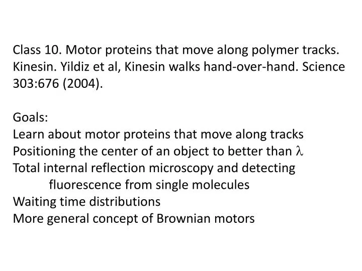
Class 10. Motor proteins that move along polymer tracks. Kinesin . Yildiz et al, Kinesin w alks hand-over-hand. Science 303:676 (2004). Goals: Learn about motor proteins that move along tracks Positioning the center of an object to better than l

E N D
Class 10. Motor proteins that move along polymer tracks. Kinesin. Yildiz et al, Kinesinwalks hand-over-hand. Science 303:676 (2004). Goals: Learn about motor proteins that move along tracks Positioning the center of an object to better than l Total internal reflection microscopy and detecting fluorescence from single molecules Waiting time distributions More general concept of Brownian motors
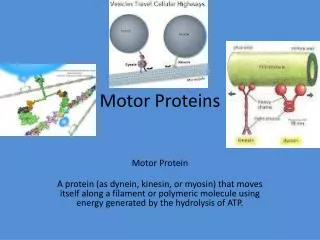
Kinesindimer cargo binding domain vesicle cargo coiled coil neck motor domain head (binds microtubule, ATP, hydrolyzes ATP) microtubule
Head groups go through cycles of binding b-tubulin and coming off One head group stays bound when other releases so kinesin stays attached through many bind/release cycles Head groups take turns binding sequential b-tubulins in protofilament so kinesin “walks” bipedially Cycles of binding and release coupled to cycles of binding and hydrolyzing ATP, releasing ADP – details unclear Interactions with tubulin, ATP, ADP etc change conformation of kinesin, which changes energy of binding other mol. Details of walk - protein conformation at each chemo- mechanical step – are subjects of great interest What makes the walk go in one direction?
Goal of paper – distinguish between 2 classes of models Strategy - label single monomer in dimer, see single label, position it to within < 8nm, measure step size
Labeling method – mutate to eliminate solvent exposed cys replace E215 or T324 or S43 with cys covalently couple fluorescent dye to mutant kinesin at dye:dimer ratio 0.4; most dimers get <1 dye dye-labeled sites (in diff. molecules) ATP binding site What would happen if both monomers got labeled?
Experimental set-up Adhere sea urchin axonemes (microtubules) to glass slide Add dye-labeled kinesins Preliminary expts – dye-labeled kinesins bind in absence of ATP but don’t move; move when ATP added
How to see fluorescence from single dye molecule Total Internal Reflection (TIR) Microscopy Limiting illumination to evanescent field markedly decreases background Sako and Uyemura, Cell Struct and Function 27:357 (2002)
How do you know you are looking at single dye molecules? Fluorescent molecules “bleach” (photo-chemical modification -> loss of fluorescence) Single step loss of intensity to 0 => single molecule ~ 400 photons/sec for 20 sec, then nothing
How do you position fluorescent spot with nm precision? Average many photons - intensity weighted position “centroid” not subject to diffraction limit ~l/2 ~6800 photons collected before bleaching (20 sec); claim ~ 3nm precision centroid = mean sdm~sd/N1/2 ~sd/30 if sd~1 pixel ~100nm sdm~0.03 pixel ~3nm
Snapshots from movie of walking kinesin dye. Pixels 86nm Amazing that what seems like fairly coarse binning does not significantly impair precision Thompson et al, Biophys J 82:2775 (2002) Valentine and Rana, IEEE Trans NuclSci 43: 2501 (1996)
Step trajectories for individual kinesin molecules with dye in different positions step wait times ~1sec => ~1000 photons/step
Step size distribution for 124 steps from 22 molecules of E215C, 12 steps from 3 molecules of T324C, and 7 steps from 1 molecule of S43C/T324C heterodimer mean +sd = 17.3 + 3.3 nm rules out inch worm model (8.3 nm steps) fits hand-over-hand model (2 x 8.3 nm)
Distribution of waiting times between steps Hand-over- hand model predicts 2 random events – What are they? Initial dip not consistent with single random event reasonable fit to P(t) = t k2 exp(-kt) (2 random events)
Check that initial dip is not instrument artifact: immobilize fluorescent beads and program stage to move 17nm at random times with average 0.6 steps/sec observed step time distribution single exponential with k = 0.65/sec
Does it make sense that the time between steps should depend on a minimum of 2 random events? Would the steps have equal rates in the inchworm model? In the hand-over-hand mechanism?
If alternate steps in hand-over-hand model were not identical due to alternately overwinding/ underwinding the dimer coiled coil, alternate waiting time distributions could be different. How could they look for this in their data? See if waiting time distribution fits two stochastic events with different waiting times: p(t) = [k1k2/(k2-k1)](e-k1t - e-k2t) Compare waiting time distributions of odd # vs even # steps: podd(t) = k1e-k1t , peven(t) = k2e-k2t
Variant hand-over-hand models: monomer detaches, pauses at relaxed position waiting for some event, e.g. ATP binding, then moves to next binding site predicts alternate steps of length x, 16.6 – x they don’t see intermediate step sizes alternate steps have same size distribution to within 2 nm 16.6 - x x
What would you expect for step size and waiting time distribution if both monomers were labeled? step size:centroid “middle” of dimer h-over-h: 8nm steps inchworm: 4nm steps waiting time: p(t) = ke-kt , since you see movement when each head steps (k1 and k2 if alt. steps differ) What would you predict for step sizes if the monomers were labeled with different color (red, green) dyes? hand-over-hand: red, green alternate16nm inchworm: each step 8nm, alternately overlapping
Brownian motor – concept underlying biomolecular motors and other nanoscale devices for directed motion simply turning on and off an asymmetric potential can -> uphill motion doesn’t violate 2nd law because takes work to turn on and off potential Asymmetric potential along “track” object that binds and diffuses when released http://www.physik.uni-augsburg.de/theo1/hanggi/Papers/309.pdf http://monet.physik.unibas.ch/~elmer/bm/ - Br. motor simulation
Applied to kinesin motor: ATP/ADP-induced conformational changes turn on and off different interactions along length of tubulin, equivalent to changing potential energy landscape Kinesin heads might usually go forward but sometimes go backward between attachments steps ATP binding/hydrolysis effect not as simple as “ATP binding or hydrolysis powers forward movement”
Conclusions Beautiful paper showing that kinesin arms move in ~17nm steps, consistent with hand-over-hand model Uses single-molecule tracking – can’t see how results could have been obtained with bulk measurements Similar methods applicable to study other motors that move along tracks – e.g. myosin on actin (in fact, authors’ previous paper was “myosin walks hand-over-hand”) Biomolecular motors suggest general principles for other nano-motors that must work in Brownian environment: alternately change binding interactions with polymer
More than half-way through course – basic ideas so far: Knowledge of structure of DNA and other biopolymers -> ways to engineer them -> novel nanostructures elastic properties of biopolymers – WLC -> ways to manipulate and measure single-molecule interactions Brownian motion in liquids at nanoscale -> diffusion, viscous drag, Brownian ratchet motors Few more examples – rotary motors, stretching proteins Think about what’s missing in understanding molecular motors and how to make new nanomachines using principles learned from biological systems