Download
1 / 26
270 likes | 479 Views
BSG Guidelines for the Diagnosis and Treatment of Cholangiocarcinoma. By Matt Johnson. Background to Guidelines. Mortality rates from intrahepatic cholangiocarcinoma have steeply risen over the course of the last 30y and continue to rise 1998+9 = 1000 deaths / year Men = Women

E N D
BSG Guidelines for the Diagnosis and Treatment of Cholangiocarcinoma By Matt Johnson
Background to Guidelines • Mortality rates from intrahepatic cholangiocarcinoma have steeply risen over the course of the last 30y and continue to rise • 1998+9 = 1000 deaths / year • Men = Women • Previously no clear national consensus for optimal diagnosis and treatment
Risk Factors • Age (65% are >65y) • PSC (lifetime risk = 5-15%) • Chronic intraductal gall stones • Bile duct adenoma + biliary papillomatosis • Caroli’s disease (cystic dilatation of ducts) • Lifetime risk = 7% • Choledochal cysts (5% will transform with time) • Smoking • Chemical Exposure ( Thorotrast, aircraft, rubber) • Tropiocal (liver flukes, chronic typhoid carriers)
Anatomical Classification • A) Intrahepatic (20-25%) • B) Perihilar (50-60%) • “Klatskin” = involve the duct bifurcation • Many are coded as intrahepatic • C) Distal Extrahepatic (20-25%) • D) Multifocal (5%)
Bismuth’s Perihilar Classification • Type 1 • Below the confluence • Type 2 • Involving the confluence but not the L or R duct • Type 3 • Occluding the CHD and involving either the L (IIIa) or the R (IIIb) hepatic duct • Type 4 • Multicentric or • Involve the CHD + both the L and the R hepatic ducts
Pathology • WHO Classification of Intrahepatic Carcinomas • Hepatocellular Ca • Combined Hepatocellular Cholangiocarcinoma • Cholangiocarcinoma, intrahepatic • Bile duct cystadenocarcinoma • Undifferentiated carcinoma
Pathology • WHO Classification of Extrahepatic bile duct carcinomas • Carcinoma in situ (* = not graded) • Adenocarcinoma (95%, graded 1-4 on glandular tissue) • Papillary adenocarcinoma (*) • Adenocarcinoma, intestinal type • Mucinous adenocarcinoma • Clear Cell adenocarcinoma (*) • Signet ring adenocarcinoma (grade 3) • Adeno-squamous carcinoma • Squamous carcinoma (graded on degree of undifferentiation) • Small cell carcinoma (“oat cell”) (grade 4) • Undifferentiated carcinoma
Molecular Diagnosis • Inactivation of tumour suppressor genes • P53, APC, Smad-4, bcl-2, p16 • Mutations in Oncogenes • K-ras, C-myc, C-erbB-2, C-neu • Chromasomal aneuploidy • 25% of perihilar tumours • These mutations can lead to phenotypic changes • Diagnostic and prognostic usefullness is unclear
Clinical Features • Obstructive Jaundice • RUQ pain, Fever + Rigors • Suggesting cholangitis • Systemic (malaise, weight loss, fatigue) • Deranged LFTs • Usually present late (esp. prox tumours)
Blood tests • Obstructive LFTs • Transaminases (us. Normal, high with acute obstruction) • Deficiency in Vit D,E,A,K (in chronic obstruction) • Chronic Systemic Markers (Hb, Alb, LDH) • CA 19-9 (85%) • Can be elevated by obstruction alone • > 100 U/ml = sensitivity of 75% and specificity of 80% • CA 125 (40-50%) • May signify the presence of peritoneal involvement) • CEA (30%)
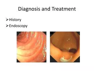
Imaging • U/S • CT + • MRI + MRCP + MRA • Cholangiography (MRCP, ERCP, PTC) • EUS • PET • Intra ductal U/S, endoscopic/percutaneous flexible cholangioscopy, radiolabelled ligand imaging
Imaging • U/S • 1st line for obstruction • Small lesions missed • Colour doppler can reveal compression/ thrombosis of the portal V or hepatic A • CT • Localises lymphadenopathy (NB. + PSC) • Enhanced spiral/ helical CT should be used if involvement of hilum or vascular system suspected • MRI • Optimal for anatomy + extent • MRCP for ductal involvement • MRA for hilar/vascular involvement
Imaging • Cholangiography • Essential for early diagnosis and assessing resectability • MRCP is non-invasive • ERCP (preferred) or PTC allows cytology and stenting • Cytology (+ in 30%) • Angiography will predict resectability NB = Biopsies should be avoided if resectability is possible
Staging • TNM or • Type 1 = limited to mucosa or muscle • Type 2 = local invasion • Type 3 = invasion of adjacent tissues or regional / hepatoD LNs (50%) • Type 4 = extensive invasion +/- distant metastases • 20% have perintoneal involvement at diagnosis
Imaging Recommendation • U/S (for initial screening) • CXR • MRI / MRCP • or contrast enhanced spiral /helical CT • Invasive Cholangiography • For tissue diagnosis +/- therapeutic decompression • Laparoscopy (if considered resectable)
Excluding Metastatic Disease • Metastatic adenocarcinoma can mimic cholangiocarcinoma • Pancreas = MRI, CT, EUS • Stomach = AXR, OGD • Breast = Ex, Mammography (if mass) • Lung = CXR • Colon = colonoscopy, spiral CT
Treatment – Curative Surgery • 5y Survival for intrahepatic Ca = 9-18% • 5y Survival for proximal Ca = 9-18% • 5y Survival for distal Ca = 20-30% • Survival depends on • stage with tumour free margins • absence of LN involvement • Median Survival • With hilar involvement = 12-24/12 • Without hilar involvement = 18-30/12
Surgery - OLTx • Contra-indicated due to rapid recurrence and death within 3y • Survival may improve in some with chemoirradiation
Palliative Procedures • Stenting • Reduces sepsis • Improves survival • Surgical bypass has not proved superior • Irradiation • Intraoperative Coeliac plexus block
Resection Reporting • 1) Tumour • Type • Extent • Blood / lymphatic involvement • Perineural invasion (worse prognosis) • 2) Margins • 3) Regional LNs (peripancreatic = distant) • 4) Additional pathological findings (PSC) • 5) Metastases
Decompression • Pre-op biliary drainage / stenting is not advised if resection being considered • May be necessary in severely malnourished or in acute suppurative cholangitis • Preop placement of biliary catheters may be a helpful technical aid when dissecting a proximal Ca
Stenting • Complex CholangioCa • MRCP will help planning management • ? Bilateral > unilateral • Plastic Vs Metal • Metal stents in those due to survive >6/12 • Metal = shorter hospital stay • Stenosis of metal stents can be treated with • Cotton-Leung plastic stent through lumen • Mesh metal stent • Semicovered stents (?reduce Ca ingrowth)
Oncology • 50% at presentation are LN+ • Treat patients early • Aim for symptom control via a multidisciplinary group as this translates into survival advantage • Aim to stabilize disease progress
Chemotherapy • Response correlates to performance status at onset • QOL is improved • No evidence to support post-surgical adjuvant therapy • No strong evidence for a survival benefit, but phase II trials suggest • Single agent 5FU partial response = 10-20% • Single agent gemcitabine partial response = 20-30% • Combination partial response = 20-40% • Gemcitabine + cisplatin partial response = 30-50% • Downstaging can convert to operability
Radiotherapy • External Beam XRT (+ChemoTx) • Palliation of painful mets + bleeding • No evidence for adjuvant post op XRT • No improvement of QOL or Survival in advanced tumours • ? Chemoradiation • Local radiation • intra-operative or intra-luminal brachytherapy • No good data to support their use • could be promising