Download
1 / 26
260 likes | 354 Views
Your Mission…. Prevention and Early Detection!!!!! Quality Improvement Evidence-based practice Teamwork and Collaboration Safety “minimize risk of harm to patients and providers through both system effectiveness and individual performance”. Indications. Long-term Caustic meds

E N D
Your Mission…. • Prevention and Early Detection!!!!! • Quality Improvement • Evidence-based practice • Teamwork and Collaboration • Safety • “minimize risk of harm to patients and providers through both system effectiveness and individual performance”
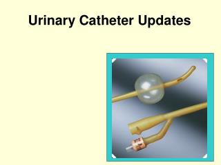
Indications • Long-term • Caustic meds • TPN (dextrose content > 10%) • Monitor RA pressures • Dialysis • Multiple therapies • No peripheral access • Frequent blood sampling
What’s in a Name? • Central Venous Access Device (CVAD) • CVC • Central line • By type (percutaneous) • TLC (triple lumen catheter), PICC • By site • subclavian, jugular, femoral • By brand name (tunneled) • Broviac, Hickman, Groshong, Mediport
What’s the Difference? Similarities • Tip of catheter in a “central” vein: • Superior vena cava Differences • How/where it is inserted • Length of stay
Method 1: Percutaneous • Needle stick, through skin, directly into vein. • Central (7 days-Phillips) • PICC (> 7 days to several months) • Single, double, or triple lumen • Triple: proximal, medial, distal ports
Tunneled • Surgical procedure • Very long-term • Exit site: chest or abdominal wall • Examples: • Hickman • Groshong • Implanted port (medi-port)
CVAD Insertion • Supplies : Check P&P • tray • antiseptic solution • Dressing material • CONSENT • 10 cc Syringes w/ NS • Needleless caps • “time out” check list
Patient Teaching r/t insertion • Purpose • Position: flat, Trendelenberg • keep hands down • face covered • turn head away
Complications of CVAD • Pneumothorax • Malposition • SVC syndrome • Occlusions • Infection • Air Embolism • Unintentional disruption
Central Venous Catheter Complications —Pneumothorax, Hemothorax, Chylothorax Cause • During insertion of CVC, introducer may cause trauma • Pneumothorax (collection of air in the pleural space due to trauma to lung) • Hemothorax (collection of blood in pleural cavity) • Chylothorax (transection of the thoracic duct causes lymph fluid to enter the pleural cavity)
Central Venous Catheter Complications: Pneumothorax Treatment • early detection: CXR after insertion • Oxygen • Monitor vital signs • Pressure should be applied over the vein entry site • Remove the catheter • Chest tube if appropriate
Obstruction – Prevention is Key • Positive Pressure Displacement device • Flush unused ports per protocol • ‘Push-Pause’ technique • Check solution for precipitates • Filter if indicated
Flushing a CVAD • 10 mL syringe or larger • Aspirate for blood return before flushing (INS,2006) • SAS or SASH (per hospital protocol) • Groshong Catheter – saline only • “push – pause” technique • Q 12 or 24 hours – per protocol • Positive pressure caps • flush, remove syringe, clamp
Infection • CRBSI • Exit site infection • Catheter tract infection • Septic thrombophlebitis
Central Venous Catheter Complications: Catheter Related Blood Stream Infection (CRBSI) Cause • Bacteria or fungi in a patient who has a intravascular device with positive blood culture • All BSIs that cannot reasonably be linked to a site of local infection are attributed to CVC • Biofilm • Contamination
Central Venous Catheter Complications:CRBSI(continued) Prevention (National Patient Safety Goals) • Strict sterile technique • Implementation of bundle approach • Tunneling and subcutaneous cuffs • Antiseptic-impregnated dressing • Colonization-resistant polymers • Contamination-resistant hubs • Luminal antimicrobial flush/lock solutions • Good hand hygiene • Frequent site assessment
CR-BSI “bundle” • Hand hygiene • Maximum barrier precautions • “time out” during insertion prn • Chlorhexidine gluconate site disinfection • Optimal catheter site (avoid femoral vein) • Daily review of line necessity – remove when no longer medically indicated.
Systemic Complication: Venous Air Embolism (VAE) Cause • Allowing the solution container to run dry and then hanging a new bag • Loose connections that allow air to enter system • Poor technique in dressing and tubing changes for central lines • Presence of air in administration set Factors that must be present: • direct communication with source of air • Pressure gradient
Systemic Complication: Venous Air Embolism (VAE) Signs and symptoms • Patient complains of palpitations • Lightheadedness and weakness • Pulmonary: dyspnea, cyanosis, tachypnea, expiratory wheezes, cough • Cardiovascular findings: “mill wheel” murmur; weak, thready pulse; tachycardia; substernal chest pain, hypotension • Neurologic findings: change in mental status, confusion, coma
Systemic Complication: Venous Air Embolism (VAE) (continued) Prevention • Purge all air from administration sets • Use 0.22 micron air-eliminating filter • Follow protocol for dressing and tubing changes for central lines • Attach piggyback meds to the proximal injection port • Use Luer-Lok connectors • Do not bypass the “pump housing” of EIDs • After removal of central lines initial dressing should be occlusive
Systemic Complication: Venous Air Embolism (VAE) (continued) Treatment • Call for help and notify physician immediately • Once VAE is suspected, any central line procedure in progress should be stopped; clamp line • Place in Trendelenburg position on left side • Administer oxygen • Maintain systemic arterial pressure with fluid resuscitation and vasopressors • Monitor vital signs • If circulatory collapse initiate CPR
CVAD Dressing Change • Prevention of infection is dependent upon • effectively reducing the number of microorganisms on the skin • Limiting access of the microorganisms to the catheter site.
Discontinuing a CVAD • Only for percutaneous • Position: Trendelenburg • Valsalva maneuver during removal • Apply pressure • Pressure dressing
Drawing blood from a central line(Dominican procedure) • Turn off IV solutions • Flush w/10 mL NS • Withdraw 5 mL “discard” • Use syringe or vacutainer to withdraw desired amt. blood • Flush w/ 20 mL NS • Label specimens “line draw”