Download
1 / 1
10 likes | 209 Views
PHANTOM FOR USE IN MR IMAGER. Missy Haehn, Can Pi, Ben Sprague, Andrea Zelisko Advisor: Professor Kristyn Masters Client: Dr. Victor Haughton. Abstract.

E N D
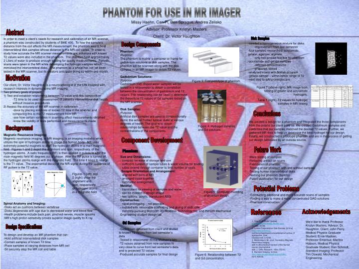
PHANTOM FOR USE IN MR IMAGER Missy Haehn, Can Pi, Ben Sprague, Andrea Zelisko Advisor: Professor Kristyn Masters Client: Dr. Victor Haughton Abstract In order to meet a client’s needs for research and calibration of an MR scanner, a phantom was constructed by students of BME 400. To how the sample’s distance from the coil affects the MR measurement, the phantom was to hold intervertebral disk samples whose distance to the MR coil varies. In order to study how accurate the MR scanner measurements are, solutions with known T2 values were also included in the phantom. The phantom held approximately 2 Liters of water to produce enough loading for quality measurements. Periodic scans were taken in the MR while developing the hydrogel samples which mimicked the intervertebral disks. The phantom as a whole has not yet been tested in the MR scanner, but its creators anticipate doing so within one month. Disk Samples -needed a homogeneous mixture for disks -improvement from last semester -four samples researched: acrylamide, gelatin, agarose, alginate -only two proved feasible for phantom -acrylamide and gelatin samples -different concentrations -prepared, tested -analyzed scans with Matlab program -gelatin sample – within better range of T2 -next step to add proteoglycans Design Components Phantom: Purpose: The phantom is mainly a container or frame for gadolinium solutions or disk samples. The phantom will be scanned along with the disk samples in an MR imager to obtain T2 values. Gadolinium Solutions: Purpose: Gadolinium (Gd) doped water samples will be tested in a relaxometer to obtain a correlation between the concentration of gadolinium and T2 values. This relationship can be used to determine the theoretical T2 values of Gd samples tested on the MR scanner. Disk Samples: Purpose: Artificial disk samples are used to compositionally mimic the actual human lumbar disks at various degrees of health. The goal is to obtain relationships between the T2 value and the concentrations of the components. Motivation Figure 3: First prototype of phantom. Our client, Dr. Victor Haughton, is a neuroradiologist at the UW-Hospital with research interests in dynamic spine MR imaging. Two primary goals of project: 1) Test and establish relationship between T2 value and disk composition -T2 time to be used in assessment of patient’s intervertebral disk health without invasive procedures 2) Assess the accuracy of the MR scanner in calibration -done by placing solutions of known T2 time in the scanner and comparing this to the T2 measurement from MR -see how certain variables in scanning affect measurements obtained -know the validity of tests performed and measurements made Figure 7 (above right): MR image from testing of gelatin and acrylamide hydrogels. Table 1 (right): T2 values for hydrogel samples in MR testing Conclusions We created a design for a phantom and integrated the three components from the criteria our client gave us. We created Gadolinium samples and confirmed that our samples matched the desired T2 values. Further, we gathered MR data to help us determine the best hydrogel for our design. Finally, we constructed a prototype phantom and are in the process of getting our design fabricated by an outside source. Figure 4: Hydrogel samples and Gd solutions. Background Magnetic Resonance Imaging: Magnetic resonance imaging, or MR imaging, is an imaging modality which utilizes the spin of hydrogen atoms to image the human body. MR uses extremely powerful magnets to align the hydrogen atoms in a main magnetic field. Figures 1 and 2 depict the alignment and spin, respectively, of the hydrogen atoms. A radio frequency (RF) is then applied in pulses to alter the main magnetic field 90 degrees out of phase. After the RF pulse is turned off, the hydrogen atoms realign with the magnetic field. The time it takes to realign is the T1 value. The exponential decay of the MR signal during the sequence of RF pulses is the T2 value. Spinal Anatomy and Imaging: -Disks act as cushions between vertebrae -Disks degenerate with age due to decreased water and blood flow -Health problems include back pain, pinched nerves, muscle spasms -MR’s high proton sensitivity proves superior image quality to X-ray Component Development Future Work Phantom • Size and Dimensions: • - compact for ease of storage and use • sufficient number of sample tubes & water volume for loading • tubes extend through container to limit number of seals • Sample Orientation and Arrangement: • - aligned with bore of MR • arranged close together • - symmetric arrangement • Material: • - translucent for viewing of samples and water • - can be molded to design shape • - two options: acrylic, translucent ABSi • Construction: • - rapid prototyping – not possible • - molded with removable scaffolding and gluing of sixth side • currently pursuing MetroRP, 3D Moldless Technology Inc. and the UW Mechanical Engineering student design lab -More testing of samples -Reducing artifact on scans -Fabrication of phantom -Testing of our phantom with and without samples -Testing human intervertebral disks -Naming our phantom (Ballots) -Patent publication for our design Figures 1 (left) and 2 (right) show the alignment and spin, respectively, of hydrogen atoms in a magnetic field. Potential Problems Figure 5: Computer modeling of phantom design. -Conducting additional and more accurate scans of samples -Finding a way to make a more concentrated GAG solutions -Phantom construction References Acknowledgements 1. “How Stuff Works” [online] http://electronics.howstuffworks.com/mri.htm. 2/1/05.2. Lumbar Degenerative Disk Disease. [online] http://www.dynomed.com/encyclopedia/encyclopedia/spine/Lumbar_Degenerative_Disk_Disease.html. 2/12/05. 3. Weidenbaum, M., et al. Correlating Magnetic Resonance Imaging with the Biochemical Content of the Normal Human Intervertebral disk. J. Ortho Research. 10(4): 552-61.4. Standard Imaging. [online] http://www.standardimaging.com/index.html. 4/20/05. We’d like to thank Professor Kristyn Masters, Advisor; Dr. Haughton, Client; John Perry, Medical Physics Graduate Student; Ernie Madsen, Professor Emeritus; Maritza Hobson, Medical Physics Graduate Student; Dan Schmidt, Standard Imaging; Professor Tim Oswald, Mechanical Engineering. Gd Samples Design Specifications -Gadolinium obtained from client and diluted to known T2 values from last semester’s data -Tested in relaxometer (Ernie Madsen's lab) -T2 values obtained from new samples fit very close to curve from last semester's data and to projected T2 values -Produced accurate samples for final design To design and develop an MR phantom that can: -Hold artificial intervertebral disk samples -Contain samples of known T2 time -Place samples at varying distances from MR coil -Sit securely atop the MR coil and table Figure 6: Relationship between T2 and Gd concentration.