Download
1 / 67
670 likes | 671 Views
Learn about the functions and types of body tissues, including epithelial, connective, muscle, and nerve tissues. Understand the characteristics and functions of each tissue type.

E N D
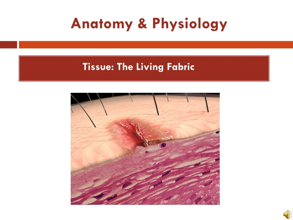
Anatomy & Physiology Tissue: The Living Fabric
Anatomy & Physiology Tissue: The Living Fabric • Objectives • Identify and describe the functions of the 4 main types of body tissues • Describe the various types and functions of epithelia • Explain the properties and functions of different types of connective tissue • Identify the major types of muscle tissue • Describe the basic types and functions of nerve tissue
Tissues • Groups of cells similar in structure and function • Types of tissues • Epithelial tissue • Connective tissue • Muscle tissue • Nerve tissue
Nervous tissue: Internal communication • Brain, spinal cord, and nerves Muscle tissue: Contracts to cause movement • Muscles attached to bones (skeletal) • Muscles of heart (cardiac) • Muscles of walls of hollow organs (smooth) Epithelial tissue: Forms boundaries between different environments, protects, secretes, absorbs, filters • Skin surface (epidermis) • Lining of GI tract organs and other hollow organs Connective tissue: Supports, protects, binds other tissues together • Bones • Tendons • Fat and other soft padding tissue Intro to tissues Figure 4.1
Epithelial Tissue (Epithelium) • Two main types (by location): • Covering and lining epithelia • On external and internal surfaces • Glandular epithelia • Secretory tissue in glands
Functions of Epithelial Tissue • 6 main functions of epithelium • Protection (skin) • Absorption (digestive tract, kidneys) • Filtration (digestive tract, kidneys) • Excretion (digestive tract, kidneys) • Secretion (glands, kidneys) • Sensory reception (skin)
Homeostatic Imbalance • Epithelial Tissue • An important characteristic of cancerous epithelial cells is their failure to respect the basement membrane boundary. (85 out of 100 cancers are of epithelial cells) • They penetrate it and invade the tissues beneath.
Characteristics of Epithelial Tissue • Cells have polarity- apical (upper, free) and basal (lower, attached) surfaces- cell regions near the apical surface differ in structure and function from cell regions in the basal surface. • Apical surfaces may have: • Microvilli: finger-like extensions of the plasma membrane that increase surface area. • Lining of the intestine • Cilia: tiny hair like projections that propel substances along their free surface. • Lining of the trachea
Characteristics of Epithelial Tissue • Are composed of closely packed cells to form continuous sheets. • Supported by a connective tissue reticular lamina (under the basal lamina) • A layer of extracellular material containing a fine network of collagen and protein fibers. • Avascular (contains no blood vessels) but innervated (supplied by nerve fibers). • Epithelial cells are nourished by substances diffusing from blood vessels in the underlying connective tissue. • High rate of regeneration • Reproduce rapidly to replace lost cells due to hostile substances in the external environment.
Classification of Epithelia Apical surface • Ask two questions: • How many layers? 1 layer = simple epithelium • Very thin most concerned with absorption, secretion, and filtration. 2 or more layers = stratified epithelium • More durable, major role is protection. Basal surface Simple Apical surface Basal surface Stratified (a) Classification based on number of cell layers. Figure 4.2a
Classification of Epithelia Squamous • What type of cell? • Squamous • Flattened and scale-like • Cuboidal • Box-like, tall as they are wide • Columnar • Tall and column shaped • (If stratified, name according to apical (top) layer of cells) Cuboidal Columnar (b) Classification based on cell shape. Figure 4.2b
Epithelia: Simple Squamous • Thin, often permeable cells • Found where filtration and exchange of substances by rapid diffusion is a priority • Kidneys • Lungs • Blood vessels
Epithelia: Simple Squamous • Two noteworthy names • Endothelium: “innercovering” slick-friction reducing lining • The lining of lymphatic vessels, blood vessels, and heart • Mesothelium: “middlecovering” • The epithelium of serous membranes in the ventral body cavity
(a) Simple squamous epithelium Description: Single layer of flattened cells with disc-shaped central nuclei and sparse cytoplasm; the simplest of the epithelia. Air sacs of lung tissue Function: Allows passage of materials by diffusion and filtration in sites where protection is not important; secretes lubricating substances in serosae. Nuclei of squamous epithelial cells Location: Kidney glomeruli; air sacs of lungs; lining of heart, blood vessels, and lymphatic vessels; lining of ventral body cavity (serosae). Photomicrograph: Simple squamous epithelium forming part of the alveolar (air sac) walls (125x). Figure 4.3a
(b) Simple cuboidal epithelium Description: Single layer of cubelike cells with large, spherical central nuclei. Simple cuboidal epithelial cells Function: Secretion and absorption. Basement membrane Location: Kidney tubules; ducts and secretory portions of small glands; ovary surface. Connective tissue Photomicrograph: Simple cuboidal epithelium in kidney tubules (430x). Figure 4.3b
(c) Simple columnar epithelium Description: Single layer of tall cells with round to oval nuclei; some cells bear cilia; layer may contain mucus- secreting unicellular glands (goblet cells). Simple columnar epithelial cell Function: Absorption; secretion of mucus, enzymes, and other substances; ciliated type propels mucus (or reproductive cells) by ciliary action. Location: Nonciliated type lines most of the digestive tract (stomach to anal canal), gallbladder, and excretory ducts of some glands; ciliated variety lines small bronchi, uterine tubes, and some regions of the uterus. Basement membrane Photomicrograph: Simple columnar epithelium of the stomach mucosa (860X). Figure 4.3c
(d) Pseudostratified columnar epithelium Description: Single layer of cells of differing heights, some not reaching the free surface; nuclei seen at different levels; may contain mucus- secreting cells and bear cilia. Cilia Mucus of mucous cell Pseudo- stratified epithelial layer Function: Secretion, particularly of mucus; propulsion of mucus by ciliary action. Location: Nonciliated type in male’s sperm-carrying ducts and ducts of large glands; ciliated variety lines the trachea, most of the upper respiratory tract. Basement membrane Photomicrograph: Pseudostratified ciliated columnar epithelium lining the human trachea (570x). Trachea Figure 4.3d
(e) Stratified squamous epithelium Description: Thick membrane composed of several cell layers; basal cells are cuboidal or columnar and metabolically active; surface cells are flattened (squamous); in the keratinized type, the surface cells are full of keratin and dead; basal cells are active in mitosis and produce the cells of the more superficial layers. Stratified squamous epithelium Function: Protects underlying tissues in areas subjected to abrasion. Nuclei Location: Nonkeratinized type forms the moist linings of the esophagus, mouth, and vagina; keratinized variety forms the epidermis of the skin, a dry membrane. Basement membrane Connective tissue Photomicrograph: Stratified squamous epithelium lining the esophagus (285x). Figure 4.3e
Epithelia: Stratified Cuboidal • Quite rare in body • Found in some sweat and mammary glands • Typically two cell layers thick Sweat Duct
Epithelia: Stratified Columnar • Limited distribution in body • Small amounts in pharynx, male urethra, and lining some glandular ducts • Also occurs at transition areas between two other types of epithelia
(f) Transitional epithelium Description: Resembles both stratified squamous and stratified cuboidal; basal cells cuboidal or columnar; surface cells dome shaped or squamouslike, depending on degree of organ stretch. Transitional epithelium Function: Stretches readily and permits distension of urinary organ by contained urine. Location: Lines the ureters, urinary bladder, and part of the urethra. Basement membrane Connective tissue Photomicrograph: Transitional epithelium lining the urinary bladder, relaxed state (360X); note the bulbous, or rounded, appearance of the cells at the surface; these cells flatten and become elongated when the bladder is filled with urine. Figure 4.3f
Answer the following questions and turn in!! • Explain what is meant by epithelial tissue being avascular but innervated. • What structure can be noted on the apical surface of the cells in this image? • What is the name of this tissue type? • A multilayered epithelium with cuboidal basal cells and flat cells at its surface would be classified as ________?
Answer the following questions and turn in!! • Explain what is meant by epithelial cells having polarity • What is significant about the cells closest to the basement membrane of this tissue type? • What is the name of this tissue type? • A multilayered epithelium with cuboidal basal cells and columnar cells at its surface would be classified as ________? • What types of organs would you find transitional epithelium in? .
Glandular Epithelia Objectives: • Define gland • Differentiate between exocrine and endocrine glands, and differentiate between multi-cellular and unicellular glands • Describe how multi-cellular exocrine glands are classified structurally and functionally
Glandular Epithelia • A gland is one or more cells that makes and secretes a particular fluid. • Glandular cells obtain needed substances from blood and transform them chemically into a product that is then released from the cell. • Classified by: • Site of product release—endocrine (internally secreting) or exocrine (externally secreting) • Relative number of cells forming the gland—unicellular (e.g., goblet cells) or multicellular (typically ducted)
Endocrine Glands • Ductless glands • Secrete hormones that travel through lymph or blood to target organs
Exocrine Glands • More numerous than endocrine glands • Secrete products into ducts • Secretions released onto body surfaces (skin) or into body cavities • Examples include mucous, sweat, oil, and salivary glands
Unicellular Exocrine Glands Microvilli Goblet Cell • The only important unicellular gland are mucous cells and goblet cells. (scattered) • Goblet cells- look like a glass with a stem due to the accumulation of mucin at the top of the cell. • Both produce mucin that dissolves in water when secreted and forms mucous- a slimy protective and lubricating coating found within the human body. Secretory vesicles containing mucin Rough ER Golgi apparatus Nucleus
Multicellular Exocrine Glands • Multicellular exocrine glands are composed of a duct and a secretory unit • Classified according to: • Duct type • Simple: unbranched duct • Compound: branched duct • Structure of their secretory units • Tubular: secretory cells form tubes. • Alveolar (acinar): secretory cells form small sacs. • Tubuloalveolar: have both types of secretory units.
Simple duct structure (duct does not branch) Compound duct structure (duct branches) Tubular secretory structure Simple tubular Simple branched tubular Compound tubular Example Intestinal glands Example Stomach (gastric) glands Example Duodenal glands of small intestine Alveolar secretory structure Simple alveolar Simple branched alveolar Compound alveolar Compound tubuloalveolar Example No important example in humans Example Sebaceous (oil) glands Example Mammary glands Example Salivary glands Surface epithelium Duct Secretory epithelium Figure 4.5
Modes of Secretion • Merocrine • Products are secreted by exocytosis • Examples: pancreas, sweat and salivary glands. • Holocrine • Products are secreted by rupture of gland cells • Example: sebaceous glands
Answer the following questions and turn in!! • What is a gland? • Explain the difference between endocrine and exocrine glands and provide an example of each • What type of an exocrine gland is a goblet cell and what does it produce? • Draw and label the various structural presentations for multicellular exocrine glands. • What is the primary difference between the way merocrine and halocrine glands secrete their products?
Connective Tissue Objectives: • Identify common characteristics of connective tissue, and list and describe its common structural elements • Describe the common types of connective tissue found in the body and indicated their particular functions
Connective Tissue • Most abundant and widely distributed tissue type • Four classes • Connective tissue proper • Cartilage • Bone tissue • Blood
Major Functions of Connective Tissue • Binding and support • Protection • Insulation • Transportation (blood)
Characteristics of Connective Tissue • Connective tissues have: • Mesenchyme (embryonic tissue) as their common tissue of origin • Varying degrees of vascularity (supply of blood vessels) • Cells separated by nonliving extracellular matrix (ground substance and fibers) • This is what enables the connective tissue to bear weight, withstand great tension, and endure physical trauma. Mesenchymal Cells Areolar Connective Tissue
Structural Elements of Connective Tissue • Ground substance • Unstructured material that fills the space between the cells and contains the fibers • Functions as a molecular sieve through which nutrients and other dissolved substances diffuse between blood capillaries and cells. • Components: • Interstitial fluid • Adhesion proteins (“glue”) • Proteoglycans • Protein core + large polysaccharides (chrondroitin sulfate and hyaluronic acid) • Trap water in varying amounts, affecting the thickness of the ground substance
Structural Elements of Connective Tissue • Fibers provide support. • Three types of fibers • Collagen (white fibers) • Strongest and most abundant type • Provides high tensile strength • Elastic • Networks of long, thin, elastin fibers that allow for stretch • Reticular • Short, fine, highly branched collagenous fibers
Structural Elements of Connective Tissue • Cells • Mitotically active and secretory cells = “blasts”= “builders” • Mature cells = “cytes” • Fibroblasts in connective tissue proper • Chondroblasts and chondrocytes in cartilage • Osteoblasts and osteocytes in bone • Hematopoietic stem cells in bone marrow • Fat cells, white blood cells, mast cells, and macrophages
Cell types Extracellular matrix Ground substance Fibers • Collagen fiber • Elastic fiber • Reticular fiber Macrophage Fibroblast Lymphocyte Fat cell Capillary Mast cell Neutrophil Figure 4.7
Connective Tissue: Embryonic • Mesenchyme—embryonic connective tissue • Gives rise to all other connective tissues • Gel-like ground substance with fibers and star-shaped mesenchymal cells
Answer the following questions and turn in!! • What are the 4 classes of connective tissue? • What 3 types of fibers provide support for connective tissue? • What are 4 functions of connective tissue? • What function(s) does adipose serve?
Connective Tissue Proper The name connective tissue proper is used to designate the connective tissue that fills interstitial spaces as opposed to the specialized connective tissues (blood, bones, cartilage, etc…). Types: • Loose connective tissue • Areolar • Adipose • Reticular • Dense connective tissue • Dense regular • Dense irregular • Elastic
(a) Connective tissue proper: loose connective tissue, areolar Description:Gel-like matrix with all three fiber types; cells: fibroblasts, macrophages, mast cells, and some white blood cells. Elastic fibers Function: Wraps and cushions organs; its macrophages phagocytize bacteria; plays important role in inflammation; holds and conveys tissue fluid. Collagen fibers Location: Widely distributed under epithelia of body, e.g., forms lamina propria of mucous membranes; packages organs; surrounds capillaries. Fibroblast nuclei Epithelium Photomicrograph: Areolar connective tissue, a soft packaging tissue of the body (300x). Lamina propria Figure 4.8a
(b) Connective tissue proper: loose connective tissue, adipose Description: Matrix as in areolar, but very sparse; closely packed adipocytes, or fat cells, have nucleus pushed to the side by large fat droplet. Function: Provides reserve food fuel; insulates against heat loss; supports and protects organs. Nucleus of fat cell Location: Under skin in the hypodermis; around kidneys and eyeballs; within abdomen; in breasts. Vacuole containing fat droplet Adipose tissue Photomicrograph: Adipose tissue from the subcutaneous layer under the skin (350x). Mammary glands Figure 4.8b
(c) Connective tissue proper: loose connective tissue, reticular Description: Network of reticular fibers in a typical loose ground substance; reticular cells lie on the network. Function: Fibers form a soft internal skeleton (stroma) that supports other cell types including white blood cells, mast cells, and macrophages. White blood cell (lymphocyte) Location: Lymphoid organs (lymph nodes, bone marrow, and spleen). Reticular fibers Spleen Photomicrograph: Dark-staining network of reticular connective tissue fibers forming the internal skeleton of the spleen (350x). Figure 4.8c
(d) Connective tissue proper: dense connective tissue, dense regular Description: Primarily parallel collagen fibers; a few elastic fibers; major cell type is the fibroblast. Collagen fibers Function: Attaches muscles to bones or to muscles; attaches bones to bones; withstands great tensile stress when pulling force is applied in one direction. Location: Tendons, most ligaments, aponeuroses. Nuclei of fibroblasts Shoulder joint Ligament Photomicrograph: Dense regular connective tissue from a tendon (500x). Tendon Figure 4.8d
(e) Connective tissue proper: dense connective tissue, dense irregular Description: Primarily irregularly arranged collagen fibers; some elastic fibers; major cell type is the fibroblast. Nuclei of fibroblasts Function: Able to withstand tension exerted in many directions; provides structural strength. Location: Fibrous capsules of organs and of joints; dermis of the skin; submucosa of digestive tract. Collagen fibers Fibrous joint capsule Photomicrograph: Dense irregular connective tissue from the dermis of the skin (400x). Figure 4.8e
(f) Connective tissue proper: dense connective tissue, elastic Description: Dense regular connective tissue containing a high proportion of elastic fibers. Function: Allows recoil of tissue following stretching; maintains pulsatile flow of blood through arteries; aids passive recoil of lungs following inspiration. Elastic fibers Location: Walls of large arteries; within certain ligaments associated with the vertebral column; within the walls of the bronchial tubes. Aorta Photomicrograph: Elastic connective tissue in the wall of the aorta (250x). Heart Figure 4.8f