Download
1 / 3
30 likes | 168 Views
Stuart S. Martin, Ph.D. Assistant Professor of Physiology University of Maryland School of Medicine HSF-2, Rm S103C 20 S. Penn St. Baltimore, MD 21201 Tel: 410-706-6601 Fax: 410-706-6600 ssmartin@som.umaryland.edu. Research Apoptosis and breast tumor metastasis

E N D
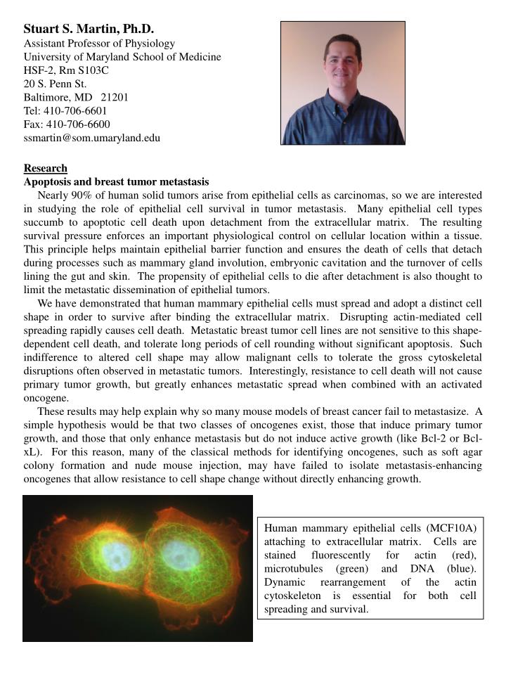
Stuart S. Martin, Ph.D. Assistant Professor of Physiology University of Maryland School of Medicine HSF-2, Rm S103C 20 S. Penn St. Baltimore, MD 21201 Tel: 410-706-6601 Fax: 410-706-6600 ssmartin@som.umaryland.edu Research Apoptosis and breast tumor metastasis Nearly 90% of human solid tumors arise from epithelial cells as carcinomas, so we are interested in studying the role of epithelial cell survival in tumor metastasis. Many epithelial cell types succumb to apoptotic cell death upon detachment from the extracellular matrix. The resulting survival pressure enforces an important physiological control on cellular location within a tissue. This principle helps maintain epithelial barrier function and ensures the death of cells that detach during processes such as mammary gland involution, embryonic cavitation and the turnover of cells lining the gut and skin. The propensity of epithelial cells to die after detachment is also thought to limit the metastatic dissemination of epithelial tumors. We have demonstrated that human mammary epithelial cells must spread and adopt a distinct cell shape in order to survive after binding the extracellular matrix. Disrupting actin-mediated cell spreading rapidly causes cell death. Metastatic breast tumor cell lines are not sensitive to this shape-dependent cell death, and tolerate long periods of cell rounding without significant apoptosis. Such indifference to altered cell shape may allow malignant cells to tolerate the gross cytoskeletal disruptions often observed in metastatic tumors. Interestingly, resistance to cell death will not cause primary tumor growth, but greatly enhances metastatic spread when combined with an activated oncogene. These results may help explain why so many mouse models of breast cancer fail to metastasize. A simple hypothesis would be that two classes of oncogenes exist, those that induce primary tumor growth, and those that only enhance metastasis but do not induce active growth (like Bcl-2 or Bcl-xL). For this reason, many of the classical methods for identifying oncogenes, such as soft agar colony formation and nude mouse injection, may have failed to isolate metastasis-enhancing oncogenes that allow resistance to cell shape change without directly enhancing growth. Human mammary epithelial cells (MCF10A) attaching to extracellular matrix. Cells are stained fluorescently for actin (red), microtubules (green) and DNA (blue). Dynamic rearrangement of the actin cytoskeleton is essential for both cell spreading and survival.
Functional genomic screening for metastatic regulators Since growth-based assays may not identify survival factors that contribute to metastatic spread, we developed an in vitro assay to select directly for resistance to shape-dependent cell death. Infection of human mammary epithelial cells with a retroviral expression library from a metastatic breast tumor cell line allowed the isolation of genes conferring a survival advantage. In addition to this genetic approach, we are using proteomics to identify new substrates of the death-associated protein kinase (DAPK). Expression of DAPK is often lost in metastatic tumor cells, but its cellular substrates remain unknown. Large-scale in vitro kinase screening of a human expressed protein array identified 15 new in vitro substrates of DAPK, and we are assessing their function in cells. Finally, we are filtering available datasets from breast cancer patients to computationally identify survival genes that may confer a higher risk of metastasis. In vivo tumor imaging In order to measure the effects of the identified genes on breast tumor metastasis, we are using in vivo imaging of transplanted tumor cells. Luciferase-expressing mouse mammary epithelial cells are used to generate cell lines stably expressing the identified survival oncogenes. Tumor growth and metastatic spread can be measured by injecting these cells into living mice and measuring the bioluminescence from the tumor cells. Since this measurement does not require sacrifice of the mice, studies addressing tumor dormancy and therapeutic response can be conducted more accurately and with far fewer mice. More importantly, since our prediction is that survival oncogenes will allow tumor spread without inducing active growth, bioluminescence allows us to follow the fate of tumor cells that fail to grow but are not eradicated. Successful treatment of such persistent and dormant tumor cells is critical for patients, since many die from recurrent metastatic disease, rather than the primary tumor. In vivo imaging of transplanted breast tumor cell lines in a single mouse. Real-time tracking and measurement of cells is possible for those tumors that grow (MEK/Bcl2) as well as those that lie dormant (EpH4 or Bcl2). At these early time points, none of the tumors are palpable, but clear differences can still be detected by bioluminescence after injection of luciferin. Personal History I received my Ph.D. from the University of California-San Diego in Biomedical Sciences, a program that combined molecular cell biology with pharmacology and physiology. As a graduate student with Dr. Jerry Olefsky, I focused on insulin-stimulated rearrangement of the actin cytoskeleton, and demonstrated that PI3-Kinase was sufficient to induce actin membrane ruffling and stress fiber breakdown. After graduating in 1998, I moved to Dr. Phil Leder's lab at Harvard Medical School to study how cell shape and the actin cytoskeleton can influence cell survival. My postdoctoral training allowed me to combine functional genomic tissue culture systems with mouse models of breast cancer. In 2004, I joined the Department of Physiology as an Assistant Professor.
Recent Publications 1. Martin, S.S., Ridgeway, A.G., Pinkas, J., Lu, Y., Reginato, M.J., Koh, E.Y., Michelman, M., Daley, G.Q., Brugge, J.S., and Leder, P. (2004). A cytoskeleton-based functional genetic screen identifies Bcl-xL as an enhancer of metastasis, but not primary tumor growth. Oncogene 23:4641-5. 2. Pinkas, J., Martin, S.S. and Leder, P. (2004) Bcl-2-mediated cell survival promotes metastasis of EpH4 bMEKDD mammary epithelial cells. Molecular Cancer Research 2:551-56. 3. Martin, S.S. and Vuori, K. (2004). Regulation of Bcl-2 proteins during anoikis and amorphosis. BBA Molecular Cell Research 1692:145-57. 4. Martin, S.S. and Leder, P. (2001). Human MCF10A mammary epithelial cells undergo apoptosis following actin depolymerization that is independent of attachment and rescued by Bcl-2. Molecular and Cellular Biology 21(19):6529-6536.