Download
1 / 44
500 likes | 1.26k Views
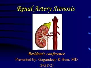
Renovascular Hypertension and Renal Artery Ultrasounds. Amajd AlMahameed, MD, MPH Division of Cardiology Beth Israel Deaconess Medical Center Boston. How Common is Renal Artery Stenosis.

E N D
Renovascular Hypertension and Renal Artery Ultrasounds Amajd AlMahameed, MD, MPH Division of Cardiology Beth Israel Deaconess Medical Center Boston
How Common is Renal Artery Stenosis Moderate RAS: visually estimated 50-69% w 10 mm Hg mean or 20 mm Hg systolic translesional gradient Patients with PAD: Significant RAS in 22% to 59% (Olin et al Am J Med. 1990;88:46N–51N, Valentine et al Ann Vasc Surg. 1993;7:220 –224.) Patients with proven history of MI: 12% had RAS > 75% (Uzu et al Am J Kidney Dis 1997;29:733-8) Severe RAS: visually estimated diameter stenosis of > 70% (Rundback et al Circulation 2002;106:1572–1585) Bilateral RAS: is not uncommon, found in 44% of RAS patients (Rimmer et al Ann Intern Med 1993;118:712-9) Necropsy Studies: Luminal stenosis > 50% was found: Overall 27-53% of autopsies Age > 70 y/o: 74% (Holley et al Am J Med. 1964;37:14 –22) (Schwartz et al BMJ. 1964;5422:1415 –1421) Patients with 1 or more clinical clues to the presence of RAS, significant RAS can be found in up to 70% (Olin et al Am J Med. 1990;88:46N–51N) CHS: significant RAS (>60%) in 6.8%, M:F ratio 2:1, W = B (Hansen et al J Vasc Surg 2002;36:443-51) White CJ. Circulation. 2006;113:1464-1473. Hirsch AT et al. Circulation 2006;113;463-654
Incidence of Renal Artery Stenosis at Cardiac Catheterization White CJ. Circulation. 2006;113:1464-1473.
Clues to RAS: Must Evaluate Such Patients White CJ. Circulation. 2006;113:1464-1473.
Renal Artery Stenosis is a PROGRESSIVE Disease Randomized trial, med Rx vs. PTA for RAS, over a 1-year, progression to RA occlusion occurred in 16% of the med Rx group compared with none in the angioplasty group (van Jaarsveld et al N Engl J Med. 2000;342:1007–1014) 29% of patients progressed, 11% developed total occlusion (mean f/u of 28 months) (Dean et al Arch Surg 1981;116:1408-15) 48% progressed from < 60% to > 60% stenosis (within 3 years) (Zierler Am J Hypertens 1996;9:1055-61) Progressive worsening of RAS occurs despite medical therapy that effectively controls blood pressure (Crowley et al Am Heart J. 1998;136:913–918, Dean et al Arch Surg. 1981;116:1408 –1415) Progression occurred at an average rate of approximately 7% per year (Zierler Am J Hypertens 1996;9:1055-61) RAS is the cause of ESRD in 15% of patients over age 50 beginning dialysis each year (Rimmer Ann Intern Med. 1993;118:712–719, Scoble et al Clin Nephrol. 1989;31:119 –122) Disease progression, based on sonographic determination, was 35% at 3 years and 51% at 5 years Caps et al Circulation 1998;98:2866-72 White CJ. Circulation. 2006;113:1464-1473. Hirsch AT et al. Circulation 2006;113;463-654
RAS: An Ominous Diagnosis to Make • RAS (50%): Stronger independent predictor of 4-yr all-cause mortality (RR 2.9) than CHF (RR 2.3), dec LVEF (RR 1.7), or dec renal function (RR 1.3) Conlon et al J Am Soc Nephrol. 1998;9:252–256 • mild-to-moderate (50%) RAS was associated with a 30% 4-year mortality rate, which almost doubled (52%) with severe (95%) RAS (incremental effect) Conlon Kidney Int. 2001;60:1490 –1497. • independent predictor of death regardless of the presence, severity, or method of revascularization of coronary artery disease Kennedy et al Am J Kidney Dis. 2003;42:926 –935, Conlon et al J Am Soc Nephrol. 1998;9:252–256, Conlon Kidney Int. 2001;60:1490 –1497. White CJ. Circulation. 2006;113:1464-1473. Hirsch AT et al. Circulation 2006;113;463-654
Indications for Revascularization • Hemodynamically significant renal artery stenosis associated with: -HTN (accelerated, resistant, malignant, with a unilaterally small kidney, and/or with intolerance to medication) - Renal insufficiency -Recurrent CHF or “flash” pulmonary edema, refractory heart failure, or refractory angina pectoris● White CJ. Circulation. 2006;113:1464-1473. Hirsch AT et al. Circulation 2006;113;463-654
Patient Selection for Renal Revascularization: Prediction of Success • Fractional flow reserve • BNP • Resistive Index White CJ. Circulation. 2006;113:1464-1473. Hirsch AT et al. Circulation 2006;113;463-654
The History of Peripheral US • Duplex scanning was introduced in 1974 • First applied to the carotid arteries • Major advances since included: ◘ Improved B-mode imaging ◘ Better low-frequency transducers (deeper penetration) ◘ Improved microprocessor software, and ◘The addition of color to B-mode image.
Provides information about native anatomy and grafts Localizes and measures stenoses Peripheral US
Identifies occlusion with reconstitution Peripheral US
Laminar Flow • Flow in a cylinder with concentric layers • Friction created between layers • Velocity is slowest near the walls and fastest in the center
Turbulent Flow • A product of FFT analysis (displays all velocities within the Doppler signal) • Spectral broadening is the term used to describe turbulent flow filling the Doppler spectral window
Turbulent Flow • Chaotic blood flow with different directions and speeds within the signal • Identified after an area of disruption of flow (severe stenosis) Turbulant flow has a shape similar to Bart Simpson’s hair!
Aliasing Aliasing corrected by dropping the baseline or increasing the scale
True Aliasing secondary to very high velocities “wrap around”
Low Cardiac Output Necessary for Standardization Proximal Lesions Same Criteria for Stents Cardiac Arrhythmia Several Limitations/Pitfalls Compensatory Flow Tortuosity Long Lesions Doppler Angle Velocity Criteria in Peripheral Ultrasounography
Hints to Proximal Stenotic Lesions • Delayed acceleration time to peak systole • Velocities alone not reliable as they may be normal, slightly elevated, or even low • Turbulence should be documented distal to the lesion • Occasionally, collateral vessels may be identified by abnormal flow patterns (retrograde) Normal Doppler signal Abnormal Doppler signal (turbulant)
Direct Assessment of RAS Velocity Criteria (+ Turbulance) Renal/Aortic Ratio Supportive findings White CJ. Circulation. 2006;113:1464-1473. Hirsch AT et al. Circulation 2006;113;463-654
Velocity Criteria for RAS • 0-59% stenosis (No clinically significant stenosis) • PSV<200cm/sec. • 60-99% stenosis (Clinically significant stenosis) • PSV>200cm/sec • Post-stenotic turbulence present • Occlusion • Artery visualized without flow White CJ. Circulation. 2006;113:1464-1473. Hirsch AT et al. Circulation 2006;113;463-654
Renal/Aortic Ratio • RAR < 3.5: Non-significant stenosis (0-59%) • RAR > 3.5: Significant stenosis (60-99%) (Sensitivity 84-88%, Specificity 97-99%, PPV 94-98%) • EDV > 150 cm/sec may indicate > 80% stenosis • Aorta velocities must be between 40 and 100cm/sec for above criteria! If not go back to PSV > 200cm/sec (and the presence of post-stenotic turbulance). Normal Renal Artery: RAR < 3.5 and PSV < 200 <60%stenosis:PSV > 200 but RAR < 3.5 (Note, AO PSV should be 40-100) 60-99% stenosis: RAR > 3.5, regardless of PSV White CJ. Circulation. 2006;113:1464-1473. Hirsch AT et al. Circulation 2006;113;463-654
PSV-EDV PSV Supportive Data (Indirect Assessment) • Resistive Index • Normal: 0.53 to 0 .70 • > 0.70: suggests intrinsic kidney disease • < 0.53: suggests renal artery stenosis • Acceleration time • Normal < 100m/sec • Tardus/parvus waveform (delayed upstroke) • Small kidney (< 8 cm in length or > 1.5 cm discrepancy from other kidney) White CJ. Circulation. 2006;113:1464-1473. Hirsch AT et al. Circulation 2006;113;463-654
R Kidney Aorta RA US showing the whole course of the R renal artery
RRA stenosis at origin. Note turbulent flow And increased velocities
Increased velocities at mid renal artery segment: Typical of non-atherosclerotic RAS (such as FMD)
RA US can evaluate the parenchymal flow as well Note increased acceleration time (AT) and borderline resistive index (RI) RI 0.53 AT 140 m/s
Another example of the common form of RAS (atherosclerotic) Lesion is typically at the origin of the vessel
RI 1.00 Example of increased resistive index (indicative Of intrinsic kidney disease)
Documentation of floe within the renal vein is part of RA US exam
Prediction of Clinical Response to Revascularization: RFFR • The renal fractional flow reserve (FFR) is an assessment of the severity of the RAS by using maximal vasodilation with papaverine • Patients with an abnormal baseline renal FFR (0.8) had a higher rate of blood pressure improvement (86%) compared with only 30% in those with a normal baseline FFR White CJ. Circulation. 2006;113:1464-1473. Mitchell J, et al. Catheter Cardiovasc Interv. 2005;65:135. Abstract.
Prediction of Clinical Response to Revascularization: BNP • Hemodynamically significant RAS activates renin-angiotensin system, leading to increased levels of angiotensin II. • In animal experiments, angiotensin II induces synthesis and release of BNP, and the BNP mRNA is upregulated in the setting of RAS. • BNP is increased in patients with refractory hypertension and renal artery stenosis • An elevated baseline BNP 80 pg/mL strongly correlated with hypertension improvement after 3.5 months of follow-up. White CJ. Circulation. 2006;113:1464-1473. Silva JA et al. Circulation. 2005;111:328 –333.
Mean (±SE) Changes in Creatinine Clearance post PTA, According to the Resistive-Index Value before Revascularization Asterisks indicate a significant difference (P<0.05) between the two groups with use of an unpaired t-test with Bonferroni's adjustment. Prediction of Clinical Response to Revascularization: BNP Radermacher et al. NEJM;344 (6): 410
Baseline One Year P >0.0001 108 P >0.05 104 P >0.0001 100 96 Mean BP 92 88 84 RI < 0.7 RI 0.7- 0.8 RI > 0.8 Mean BP in patients without nephrosclerosis (RI <0.7), with mild nephrosclerosis (RI, 0.7 to 0.8), and with severe nephrosclerosis (RI >0.8) White CJ. Circulation. 2006;113:1464-1473. Zelelr T et al. Circulation. 2003;108:2244 –2249.
Baseline One Year P < 0.05 P < 0.05 P = NS Serum Cr Serum Cr in patients without nephrosclerosis (RI <0.7), mild nephrosclerosis (RI, 0.7 to 0.8), and severe nephrosclerosis (RI >0.8) RI < 0.7 RI 0.7-0.8 RI > 0.8 White CJ. Circulation. 2006;113:1464-1473. Zelelr T et al. Circulation. 2003;108:2244 –2249.