Download
1 / 1
10 likes | 165 Views
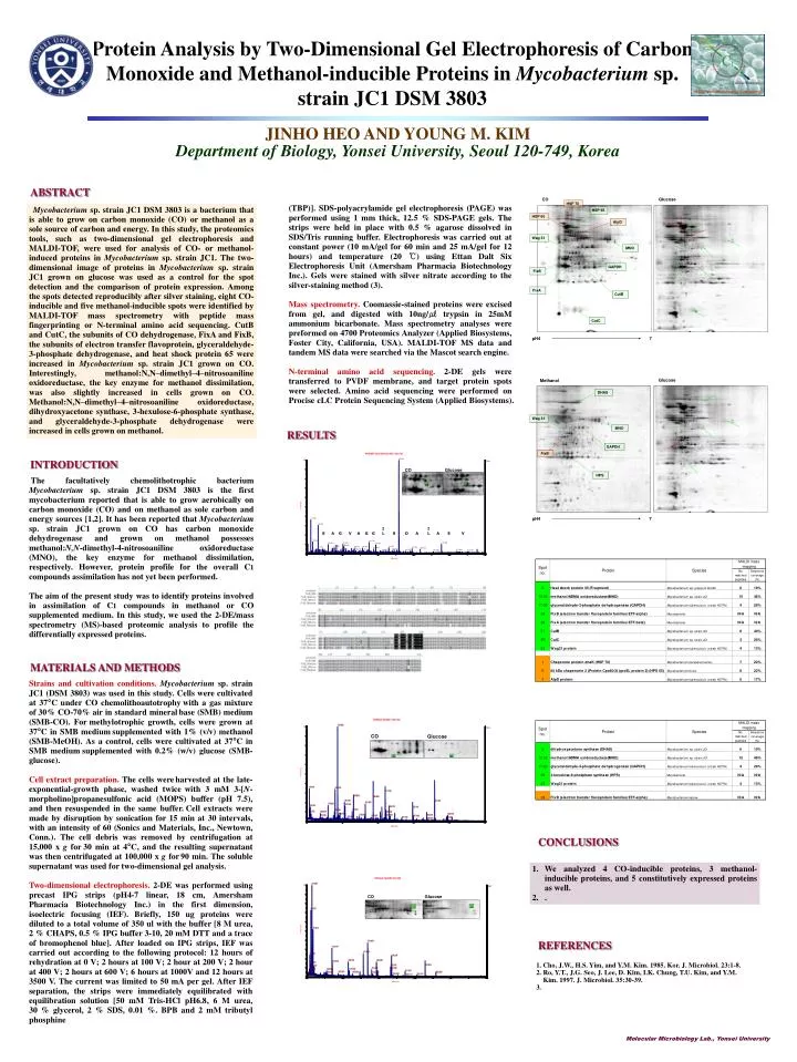
CO. Glucose. Glucose. Methanol. HSP 70. HSP 65. DHAS. HSP 60. AtpD. 31. 31. Wag 31. Wag 31. MNO. MNO. GAPDH. GAPDH. FixB. FixB. Fix A. CutB. HPS. I L. I L. CutC. S. A. G. V. A. G. G. S. D. A. A. E. V. 7. 7. pH4. pH4.

E N D
CO Glucose Glucose Methanol HSP 70 HSP 65 DHAS HSP 60 AtpD 31 31 Wag 31 Wag 31 MNO MNO GAPDH GAPDH FixB FixB FixA CutB HPS I L I L CutC S A G V A G G S D A A E V 7 7 pH4 pH4 Protein Analysis by Two-Dimensional Gel Electrophoresis of Carbon Monoxide and Methanol-inducible Proteins in Mycobacterium sp. strain JC1 DSM 3803 JINHO HEO AND YOUNG M. KIM Department of Biology, Yonsei University, Seoul 120-749, Korea ABSTRACT (TBP)]. SDS-polyacrylamide gel electrophoresis (PAGE) was performed using 1 mm thick, 12.5 % SDS-PAGE gels. The strips were held in place with 0.5 % agarose dissolved in SDS/Tris running buffer. Electrophoresis was carried out at constant power (10 mA/gel for 60 min and 25 mA/gel for 12 hours) and temperature (20 ℃) using Ettan Dalt Six Electrophoresis Unit (Amersham Pharmacia Biotechnology Inc.). Gels were stained with silver nitrate according to the silver-staining method (3). Mass spectrometry. Coomassie-stained proteins were excised from gel, and digested with 10ng/㎕ trypsin in 25mM ammonium bicarbonate. Mass spectrometry analyses were preformed on 4700 Proteomics Analyzer (Applied Biosystems, Foster City, California, USA). MALDI-TOF MS data and tandem MS data were searched via the Mascot search engine. N-terminal amino acid sequencing. 2-DE gels were transferred to PVDF membrane, and target protein spots were selected. Amino acid sequencing were performed on Procise cLC Protein Sequencing System (Applied Biosystems). Mycobacterium sp. strain JC1 DSM 3803 is a bacterium that is able to grow on carbon monoxide (CO) or methanol as a sole source of carbon and energy. In this study, the proteomics tools, such as two-dimensional gel electrophoresis and MALDI-TOF, were used for analysis of CO- or methanol-induced proteins in Mycobacterium sp. strain JC1. The two-dimensional image of proteins in Mycobacterium sp. strain JC1 grown on glucose was used as a control for the spot detection and the comparison of protein expression. Among the spots detected reproducibly after silver staining, eight CO-inducible and five methanol-inducible spots were identified by MALDI-TOF mass spectrometry with peptide mass fingerprinting or N-terminal amino acid sequencing. CutB and CutC, the subunits of CO dehydrogenase, FixA and FixB, the subunits of electron transfer flavoprotein, glyceraldehyde-3-phosphate dehydrogenase, and heat shock protein 65 were increased in Mycobacterium sp. strain JC1 grown on CO. Interestingly, methanol:N,N–dimethyl–4–nitrosoaniline oxidoreductase, the key enzyme for methanol dissimilation, was also slightly increased in cells grown on CO. Methanol:N,N–dimethyl–4–nitrosoaniline oxidoreductase, dihydroxyacetone synthase, 3-hexulose-6-phosphate synthase, and glyceraldehyde-3-phosphate dehydrogenase were increased in cells grown on methanol. RESULTS INTRODUCTION CO Glucose The facultatively chemolithotrophic bacterium Mycobacterium sp. strain JC1 DSM 3803 is the first mycobacterium reported that is able to grow aerobically on carbon monoxide (CO) and on methanol as sole carbon and energy sources [1,2]. It has been reported that Mycobacterium sp. strain JC1 grown on CO has carbon monoxide dehydrogenase and grown on methanol possesses methanol:N,N-dimethyl-4-nitrosoaniline oxidoreductase (MNO), the key enzyme for methanol dissimilation, respectively. However, protein profile for the overall C1 compounds assimilation has not yet been performed. The aim of the present study was to identify proteins involved in assimilation of C1 compounds in methanol or CO supplemented medium. In this study, we used the 2-DE/mass spectrometry (MS)-based proteomic analysis to profile the differentially expressed proteins. 24 24 MATERIALS AND METHODS Strains and cultivation conditions.Mycobacterium sp. strain JC1 (DSM 3803) was used in this study. Cells were cultivated at 37°C under CO chemolithoautotrophywith a gas mixture of 30% CO-70% air in standard mineralbase (SMB) medium (SMB-CO). Formethylotrophic growth, cells were grown at 37°C in SMB mediumsupplemented with 1% (v/v) methanol (SMB-MeOH). As a control, cells were cultivated at 37°C in SMB mediumsupplemented with 0.2% (w/v) glucose (SMB-glucose). Cell extract preparation. The cells wereharvested at the late-exponential-growth phase, washed twicewith 3 mM 3-[N-morpholino]propanesulfonic acid (MOPS) buffer (pH 7.5), and then resuspended in the same buffer.Cell extracts were made by disruption by sonication for 15 min at 30 intervals, with an intensity of 60 (Sonics and Materials, Inc., Newtown, Conn.). The cell debris was removed by centrifugation at 15,000 x g for30 min at 4°C, and the resulting supernatant was then centrifugated at 100,000 x g for90 min. The soluble supernatant was used for two-dimensional gel analysis. Two-dimensional electrophoresis. 2-DE was performed using precast IPG strips (pH4-7 linear, 18 cm, Amersham Pharmacia Biotechnology Inc.) in the first dimension, isoelectric focusing (IEF). Briefly, 150 ug proteins were diluted to a total volume of 350 ul with the buffer [8 M urea, 2 % CHAPS, 0.5 % IPG buffer 3-10, 20 mM DTT and a trace of bromophenol blue]. After loaded on IPG strips, IEF was carried out according to the following protocol: 12 hours of rehydration at 0 V; 2 hours at 100 V; 2 hour at 200 V; 2 hour at 400 V; 2 hours at 600 V; 6 hours at 1000V and 12 hours at 3500 V. The current was limited to 50 mA per gel. After IEF separation, the strips were immediately equilibrated with equilibration solution [50 mM Tris-HCl pH6.8, 6 M urea, 30 % glycerol, 2 % SDS, 0.01 %. BPB and 2 mM tributyl phosphine CO Glucose CONCLUSIONS • We analyzed 4 CO-inducible proteins, 3 methanol-inducible proteins, and 5 constitutively expressed proteins as well. • . CO Glucose 11 11 REFERENCES 1. Cho, J.W., H.S. Yim, and Y.M. Kim. 1985. Kor. J. Microbiol. 23:1-8. 2. Ro, Y.T., J.G. Seo, J. Lee, D. Kim, I.K. Chung, T.U. Kim, and Y.M. Kim. 1997. J. Microbiol. 35:30-39. 3. Molecular Microbiology Lab., Yonsei University