Download
1 / 40
430 likes | 577 Views
© 2012 Pearson Education, Inc. 11.1 Restriction and Modification Enzymes. Genetic engineering : using in vitro techniques to alter genetic material in the laboratory Basic techniques include Restriction enzymes Gel electrophoresis Nucleic acid hybridization Nucleic acid probes

E N D
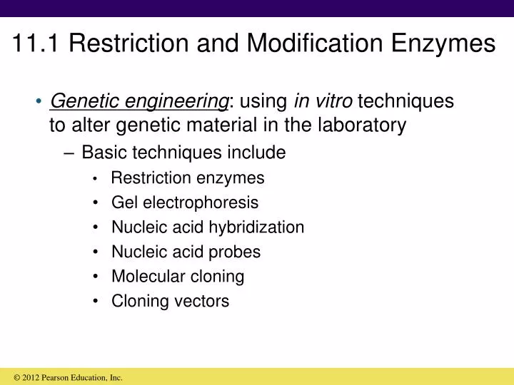
© 2012 Pearson Education, Inc. 11.1 Restriction and Modification Enzymes • Genetic engineering: using in vitro techniques to alter genetic material in the laboratory • Basic techniques include • Restriction enzymes • Gel electrophoresis • Nucleic acid hybridization • Nucleic acid probes • Molecular cloning • Cloning vectors
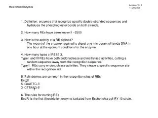
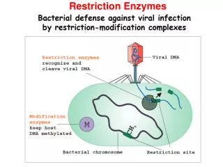
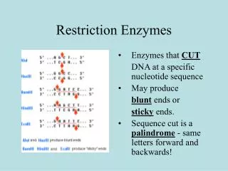
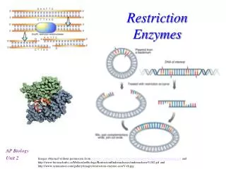
© 2012 Pearson Education, Inc. 11.1 Restriction and Modification Enzymes • Restriction enzymes: recognize specific DNA sequences and cut DNA at those sites • Widespread among prokaryotes • Rare in eukaryotes • Protect prokaryotes from hostile foreign DNA (e.g., viral genomes) • Essential for in vitro DNA manipulation
© 2012 Pearson Education, Inc. 11.1 Restriction and Modification Enzymes • Three classes of restriction enzymes • Type II cleave DNA within their recognition sequence and are most useful for specific DNA manipulation (Figure 11.1a) • Restriction enzymes recognize inverted repeat sequences (palindromes) • Typically 4–8 base pairs long; EcoRI recognizes a 6-base-pair sequence • Sticky ends or blunt ends
Figure 11.1a Single-stranded“sticky” ends © 2012 Pearson Education, Inc.
© 2012 Pearson Education, Inc. 11.1 Restriction and Modification Enzymes • Restriction enzymes protect cell from invasion from foreign DNA • Destroy foreign DNA • Must protect their own DNA from inadvertent destruction
© 2012 Pearson Education, Inc. 11.1 Restriction and Modification Enzymes • Modification enzymes: protect cell’s DNA for restriction enzymes • Chemically modify nucleotides in restriction recognition sequence • Modification generally consists of methylation of DNA (Figure 11.1b)
Figure 11.1b © 2012 Pearson Education, Inc.
© 2012 Pearson Education, Inc. 11.1 Restriction and Modification Enzymes • Gel electrophoresis: separates DNA molecules based on size (Figure 11.2a) • Electrophoresis uses an electrical field to separate charged molecules • Gels are usually made of agarose, a polysaccharide • Nucleic acids migrate through gel toward the positive electrode due to their negatively charged phosphate groups • Gels can be stained with ethidium bromide and DNA can be visualized under UV light (Figure 11.2b)
Figure 11.2a © 2012 Pearson Education, Inc.
B C D A Size in basepairs Size in basepairs 5000 4000 3000 Figure 11.2b 2000 1800 1000 500 © 2012 Pearson Education, Inc.
© 2012 Pearson Education, Inc. 11.1 Restriction and Modification Enzymes • The same DNA that has been cut with different restriction enzymes will have different banding patterns on an agarose gel • Size of fragments can be determined by comparison to a standard • Restriction map: a map of the location of restriction enzyme cuts on a segment of DNA (Figure 11.3)
© 2012 Pearson Education, Inc. 11.2 Nucleic Acid Hybridization • Nucleic acid hybridization: base pairing of single strands of DNA or RNA from two different sources to give a hybrid double helix • Segment of single-stranded DNA that is used in hybridization and has a predetermined identity is called a nucleic acid probe • Southern blot: a hybridization procedure where DNA is in the gel and probe is RNA or DNA • Northern blot: RNA is in the gel
Figure 11.4 © 2012 Pearson Education, Inc.
© 2012 Pearson Education, Inc. 11.3 Essentials of Molecular Cloning • Molecular cloning: isolation and incorporation of a piece of DNA into a vector so it can be replicated and manipulated • Three main steps of gene cloning (Figure 11.5): • Isolation and fragmentation of source DNA • Insertion of DNA fragment into cloning vector • Introduction of cloned DNA into host organism
Foreign DNA Cut with restrictionenzyme Figure 11.5 Add vector cutwith same restriction enzyme Stickyends Vector Add DNA ligase toform recombinantmolecules ClonedDNA Introduction of recombinantvector into a host © 2012 Pearson Education, Inc.
© 2012 Pearson Education, Inc. 11.3 Essentials of Molecular Cloning • Isolation and fragmentation of source DNA • Source DNA can be genomic DNA, RNA, or PCR-amplified fragments • Genomic DNA must first be restriction digested
© 2012 Pearson Education, Inc. 11.3 Essentials of Molecular Cloning • Insertion of DNA fragment into cloning vector • Most vectors are derived from plasmids or viruses • DNA is generally inserted in vitro • DNA ligase: enzyme that joins two DNA molecules • Works with sticky or blunt ends
© 2012 Pearson Education, Inc. 11.3 Essentials of Molecular Cloning • Introduction of cloned DNA into host organism • Transformation is often used to get recombinant DNA into host • Some cells will contain desired cloned gene, while other cells will have other cloned genes • Gene library: mixture of cells containing a variety of genes • Shotgun cloning: gene libraries made by cloning random genome fragments Animation: Recombinant DNA
© 2012 Pearson Education, Inc. 11.3 Essentials of Molecular Cloning • Essential to detect the correct clone • Initial screen: antibiotic resistance, plaque formation • Often sufficient for cloning of PCR-generated DNA sequences • If working with a heterogeneous gene library you may need to look more closely
Transformant coloniesgrowing on agar surface Replica-plate ontomembrane filter Figure 11.6 Lyse bacteria and denatureDNA; add RNA or DNAprobe (radioactive); washout unbound radioactivity Partially lyse cells; addspecific antibody; add agentto detect bound antibody inradiolabeled form Autoradiographto detectradioactivity X-ray film Positivecolonies © 2012 Pearson Education, Inc.
© 2012 Pearson Education, Inc. 11.4 Molecular Methods for Mutagenesis • Synthetic DNA • Systems are available for de novo synthesis of DNA • Oligonucleotides of 100 bases can be made • Multiple oligonucleotides can be ligated together • Synthesized DNA is used for primers and probes, and in site-directed mutagenesis
© 2012 Pearson Education, Inc. 11.4 Molecular Methods for Mutagenesis • Conventional mutagens produce mutations at random • Site-directed mutagenesis: performed in vitro and introduces mutations at a precise location (Figure 11.7) • Can be used to assess the activity of specific amino acids in a protein • Structural biologists have gained significant insight using this tool
Clone into single-strandedvector Source Single-strandedDNA from M13phage Add syntheticoligonucleotidewith one basemismatch Base-pairingwith sourcegene Figure 11.7 Extend singlestrand withDNA polymerase Transformationand selection Clone andselect mutant © 2012 Pearson Education, Inc.
© 2012 Pearson Education, Inc. 11.4 Molecular Methods for Mutagenesis • Cassette mutagenesis and knockout mutations • DNA fragment can be cut, excised, and replaced by a synthetic DNA fragment (DNA cassettes or cartridges) • The process is known as cassette mutagenesis • Gene disruptionis when cassettes are inserted into the middle of the gene (Figure 11.8) • Gene disruption causes knockout mutations
EcoRI cut sites () Gene X Kanamycin cassette Cut with EcoRIand ligate Figure 11.8 BamHIcut site Cut with BamHI andtransform into cellwith wild-type gene X Linearized plasmid Sites of recombination Chromosome Recombination and selectionfor kanamycin-resistant cells Gene X knockout © 2012 Pearson Education, Inc.
Figure 11.9 © 2012 Pearson Education, Inc.
Target gene Coding sequence Promoter Reporter gene Figure 11.10 Promoter Coding sequence Cut and ligate Gene fusion Promoter Reporter is expressed undercontrol of target gene promoter Reporterenzyme Substrate Colored product © 2012 Pearson Education, Inc.
© 2012 Pearson Education, Inc. 11.6 Plasmids as Cloning Vectors • Plasmids are natural vectors and have useful properties as cloning vectors • Small size; easy to isolate DNA • Independent origin of replication • Multiple copy number; get multiple copies of cloned gene per cell • Presence of selectable markers • Vector transfer carried out by chemical transformation or electroporation
© 2012 Pearson Education, Inc. 11.6 Plasmids as Cloning Vectors • pUC19 is a common cloning vector (Figure 11.11) • Modified ColE1 plasmid • Contains ampicillin resistance and lacZ genes • Contains polylinker (multiple cloning site) within lacZ gene
Order of restrictionenzyme cut sites inpolylinker Ampicillinresistance lacZ ApoI - EcoRIBanII - SacIAcc651 - KpnIAvaI - BsoBI -SmaI - XmaIBamHIXbaIAccI - HincII - SalIBspMI - BfuAISbfIPstISphIHindIII Figure 11.11 Polylinker pUC192686 base pairs lacI Origin ofDNA replication © 2012 Pearson Education, Inc.
© 2012 Pearson Education, Inc. 11.6 Plasmids as Cloning Vectors • Blue/white screening • Blue colonies do not have vector with foreign DNA inserted • White colonies have foreign DNA inserted • Insertional inactivation: lacZ gene is inactivated by insertion of foreign DNA (Figure 11.12) • Inactivated lacZ cannot process Xgal; blue color does not develop
lacZ AmpR Foreign DNA Vector Digestion with restriction enzyme Figure 11.12 Join withDNA ligase Opened vector Recyclized vector without insert Vector plus foreignDNA insert Transform into Escherichiacoli and select on ampicillinplates containing Xgal Transformants blue(-galactosidaseactive) Transformants white(-galactosidaseinactive) © 2012 Pearson Education, Inc.
© 2012 Pearson Education, Inc. 11.7 Hosts for Cloning Vectors • Ideal hosts should be • Capable of rapid growth in inexpensive medium • Nonpathogenic • Capable of incorporating DNA • Genetically stable in culture • Equipped with appropriate enzymes to allow replication of the vector • Escherichia coli, Bacillus subtilis, Saccharomyces cerevisiae
Eukaryote Bacteria Escherichia coli Bacillus subtilis Saccharomycescerevisiae Figure 11.13 Easily transformedNonpathogenicNaturally secretes proteinsEndospore formation simplifies culture Well-developed geneticsNonpathogenicCan process mRNA and proteinsEasy to grow Well-developed geneticsMany strains availableBest known bacterium Genetically unstableGenetics less developed than in E. coli Potentially pathogenicPeriplasm traps proteins Plasmids unstableWill not replicate most bacterial plasmids Advantages Disadvantages © 2012 Pearson Education, Inc.
© 2012 Pearson Education, Inc. 11.5 Gene Fusions and Reporter Genes • Reporter genes • Encode proteins that are easy to detect and assay (Figure 11.9) • Examples:lacZ, luciferase, GFP genes • Gene fusions • Promoters or coding sequences of genes of interest can be swapped with those of reporter genes to elucidate gene regulation under various conditions(Figure 11.10)
© 2012 Pearson Education, Inc. 11.8 Shuttle Vectors and Expression Vectors • Expression vectors: allow experimenter to control the expression of cloned genes (Figure 11.16) • Based on transcriptional control • Allow for high levels of protein expression • Strong promoters • lac, trp, tac, trc, lambda PL • Effective transcription terminators are used to prevent expression of other genes on the plasmid
trc promoter lacO S/D lacI Polylinker(cloningsite) Figure 11.16 T1 T2 Ampicillinresistance Origin ofDNA replication © 2012 Pearson Education, Inc.
© 2012 Pearson Education, Inc. 11.8 Shuttle Vectors and Expression Vectors • In T7 expression vectors, cloned genes are placed under control of the T7 promoter (Figure 11.17) • Gene for T7 RNA polymerase present and under control of easily regulated system (e.g., lac) • T7 RNA polymerase recognizes only T7 promoters • Transcribes only cloned genes • Shuts down host transcription
Induce lacpromoter withIPTG T7 RNApolymerase Geneproduct Figure 11.17 lacoperator Gene forT7 RNApolymerase lacpromoter T7promoter Cloned gene pET plasmid Chromosome lacl © 2012 Pearson Education, Inc.
© 2012 Pearson Education, Inc. 11.8 Shuttle Vectors and Expression Vectors • mRNA produced must be efficiently translated and there are problems with this always happening • Bacterial ribosome binding sites are not present in eukaryotic genomes • Differences in codon usage between organisms • Eukaryotic genes containing introns will not be expressed properly in prokaryotes