Download
1 / 29
320 likes | 510 Views
14 STEPS TO ASSURE A SUCCESSFUL READING AND UNDERSTANDING OF AN UNKNOWN ECG

E N D
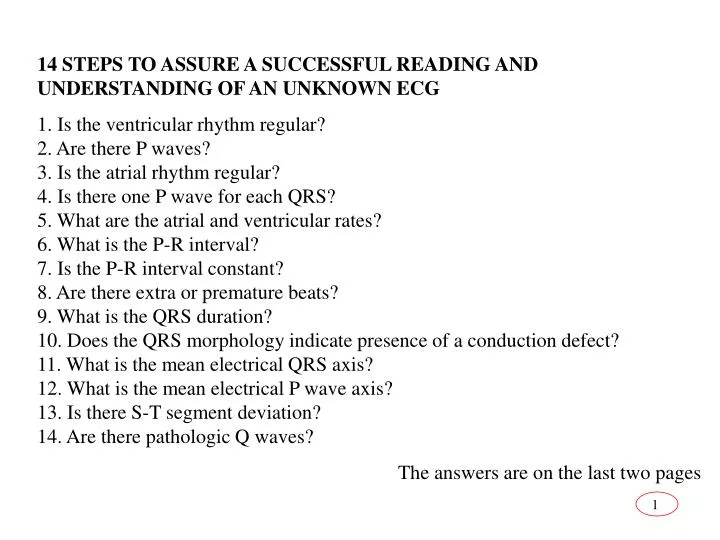
14 STEPS TO ASSURE A SUCCESSFUL READING AND UNDERSTANDING OF AN UNKNOWN ECG 1. Is the ventricular rhythm regular?2. Are there P waves?3. Is the atrial rhythm regular?4. Is there one P wave for each QRS?5. What are the atrial and ventricular rates?6. What is the P-R interval?7. Is the P-R interval constant?8. Are there extra or premature beats?9. What is the QRS duration?10. Does the QRS morphology indicate presence of a conduction defect?11. What is the mean electrical QRS axis?12. What is the mean electrical P wave axis?13. Is there S-T segment deviation?14. Are there pathologic Q waves? The answers are on the last two pages
Key to the ECGs 2. right bundle branch block plus atrial fibrillation 3. Ischemia of the bottom of the heart (inferior infarction) 4. right bundle branch block 5. first degree heart block 6. complete heart block 7. ventricular tachycardia 8. WPW 9. atrial fibrillation 10. Mobitz I second degree heart block 11. PVC’s (unifocal) 12. normal sinus rhythm 13. complete heart block (third degree) 14. second degree heart block (2:1 rhythm) 15. atrial fibrillation
16. PVC’s (unifocal) 17. normal sinus rhythm 18. sinus bradycardia 19. sinus tachycardia 20. PVC showing its +90º axis 21. Multifocal PVCs 22. atrial fibrillation with a slow ventricular rate 23. a short run of ventricular tachycardia (must be 3 or more to be considered VT, otherwise they would just be called multiple PVCs) 24. ventricular fibrillation 25. ventricular tachycardia 26. WPW 27. Mobitz I heart block. It also shows ST depression probably from ischemia on a region of the heart away from this lead