Download
1 / 53
530 likes | 733 Views
Molecular aspects of cell death. Aleksander L. Sieroń Department M olecular biology a nd genetics http://biolmolgen.slam.katowice.pl. CYCLIN. Cdk. ADP. pRB : E2F. E2F. ppRB. p53. MDM4. DNA damage. MDM2 p53 stabilization. p21 WAF1/CIP. Modified from. CY C LIN : Cdk. ATP.

E N D
Molecular aspects of cell death Aleksander L. Sieroń Department Molecular biologyand genetics http://biolmolgen.slam.katowice.pl
CYCLIN Cdk ADP pRB : E2F E2F ppRB p53 MDM4 DNA damage MDM2 p53 stabilization p21WAF1/CIP Modified from CYCLIN : Cdk ATP pRB : E2F PHASE G1 PHASE S PHASE S PHASE G1 REPAIR OR APOPTOSIS
CYCLIN : Cdk ATP pRB : E2F CYCLIN Cdk ADP pRB : E2F PHASE G1 PHASE S E2F ppRB PHASE S PHASE G1 REPAIR OR APOPTOSIS LACK OF p53 ACTIVITY
CLASSIFICATION OF MEDULOBLASOMA Classical meduloblastoma Neuroblasticmeduloblastoma Desmoplasticmeduloblastoma Kryńska B., et al. (1999) PNAS, 96:11519-24
Arrows point to cells containing JCV T-antigen Arrow heads point to proliferating cells Magnifications x400 (A & B); x200 (C); x1000 (wstawka) IMMUNOHISTOCHEMICAL DETECTION OF JCV T-ANTIGEN IN MEDULOBLASTOMS Kryńska B., et al. (1999) PNAS, 96:11519-24
Synaptophysin btubulinclassIII Magnificationsx200 (D) & x400 (E i F). GFAP IMMUNOHISTOCHEMICAL ANALYSIS OF MEDULOBLASTOMA MARKERS Kryńska B., et al. (1999) PNAS, 96:11519-24
Molecular ResearchDetection of pathogenicDNA(diagnosis based on the analysis of PCR products)
5130/0 Control region (4408-4427) 5013 (4303-4327) 277 4771 Agno 173 bp t 492 4426 526 4495 883 (4255-4274) 0.6 0.7 VP2 0.8 0.5 VP3 JCV (Mad-1) 5130 bp 0.4 0.9 1469 0.3 0.0 1560 0.1 0.2 T (1848-1828) VP1 2533 2603 212 bp (2039-2019) (1891-1872) (2797-2776) (2600-2578) 220 bp PCRprimers DNAtemplate Probe (2668-2663) GENOME of JCV Kryńska B., et al. (1999) PNAS, 96:11519-24
N-terminal coding early gene region of T-antigen T C-terminal early gene region of T-antigen Late region genes coding VP1 capsid protein DETECTION of JCV DNA by PCR METHOD IN MEDULOBLASTOMAS Kryńska B., et al. (1999) PNAS, 96:11519-24
Molecular ResearchDetecting the presence and activityof cell cycle regulators.
Immuno-co-precipitation T-antigen & p53 protein complexes Kryńska B., et al. (1997) JCB, 67:223-30
p21 EXPRESSION IN THE T-ANTIGEN PRESENCE Kryńska B., et al. (1997) JCB, 67:223-30
CYCLINS E and A and CDK2 ExPRESSION IN THE T-ANTIGEN PRESENCE Kryńska B., et al. (1997) JCB, 67:223-30
CYCLIN D, CDK4, & CDK6 EXPRESSION IN THE T-ANTIGEN PRESENCE Kryńska B., et al. (1997) JCB, 67:223-30
BLOCKING OF APOPTOSIS BY p300 REQUIRES PRESENCE OF CYCLIN D1
Immuno-co-precipitation T-ANTIGEN & pRB COMPLEXES Kryńska B., et al. (1997) JCB, 67:223-30
E2F & PCNA EXPRESSION IN TEH T-ANTIGEN PRESENCE PCNA – ProliferationCellsNuclearAntigen Kryńska B., et al. (1997) JCB, 67:223-30
Mutation Mutation Mutation Single p53 allele mutations Homologous recombination de Vries A. et al. (2002) PNAS, 99:2948-53.
Both p53 alleles inactive Mutation p53 heterozygotic One p53 allele inactive Normal/ wild p53 allele Effect of single allele of p53 gene mutations On embryonic cell survival de Vries A. et al. (2002) PNAS, 99:2948-53.
Wpływ mutacji pojedynczego allelu genu p53 na przeżywalność komórek grasicy p53 dependent apoptosis p53 independent apoptosis de Vries A. et al. (2002) PNAS, 99:2948-53.
1–5, Nature Genetics,FEBRUARY 11, 2002,online publication Ronit I. Yarden1, Sherly Prado-Reoyo1, Magda Sgagias2, Kenneth H. Cowan2 & Lowrence C. Brody1 1 Genome Technology Branch, National HumanGenomeResearchInstitute, National Institutes of Health, Bethesda, MD 2 EppleyInstitute of CancerResearch, University of Nebraska Medical Center, NA BRCA1 regulatesthe G2/M checkpoint by activating Chk1 kinase upon DNA damage Doesitexplains GENESIS OF FAMILIAL BREAST AND OVARY CENCER?
DNA DAMAGE ATM/ATR (ataxia telangiectasia mutated/ATM and Rad3-related) ATM, ATR & hCds1/Chk2 proteins of DNA damageresponse changing BRCA phosphorylation ? BRCA1 hCds1/Chk2 Chk1 – regulatory kinase Wee1 Kinase Cdc25C Cdk1/cyclinB G2 M
DNA Damage ATM/ATR ATM, ATR & hCds1/Chk2 proteins of DNA damage response changing BRCA phosphorylation ? BRCA1 hCds1/Chk2 Chk1 – Regulatory kinase Wee1 Kinase Cdc25C Cdk1/cyclin B G2 M M U T A T I O N S UNCONTROLED PROLIFERATION
BRCA1 as a suppressor of breast and ovarian tumors growth (specificity of tumor growth in heterozygous patients relative to the mutations knocking off BRCA1 activity LOH: loss of heterozygositycolored circles indicate cells with defined genotypeschange the color wheel means a change in the genotype of cells LOH: loss of heterozygositycolored circles indicate cells with defined genotypes change of the circlecolor means a change in the genotype of cells.
The dotted lines (crosses) indicate defects in pathways ATM and/or ATR occur in different cancer cell lines.
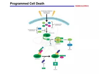
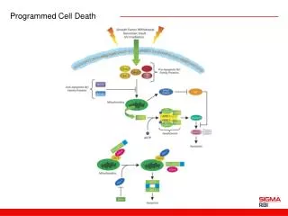
DNA damage Shortening Telomeres Oncogenes Model of apoptotic response of cells tocytotoxins, radiation and shortage of cytokines. BH3-only proteins Puma, Noxa and Bim mediate in both dependent and independent of p53 response to stress. These responses are activated equally by stress (shortage of cytokines, cytotoxins and radiation) as well as of stress signals, such as those produced during the tumorigenesis of activated oncogenes the loss of telomere and hypoxia.Activation of the ATM kinase or Arf blocks MDM2 protein, which causes the increase of p53 concentration in cells. The p53 induces the transcription of genes p21, Puma and Noxa, and of other poorly understood genes. Activating Bcl-2 (and other proteins related to it) for BH3-only proteins results in activation of Bax and Bak that induces apoptosis.Abbreviations: STS, staurosporine, dex, dexamethasone. ATM Arf Mdm2 ? Hypoxy ? p53 Lack of cytokines Ca2+ efflux p21 PMA Taxol Cell cycle arrest STS dex PIG3, PIG8, PERP P53AIP1, p53DINP1 Noxa Puma Bim ? Bcl-2 Bax/ Bak Apoptosis Villunger A. et al. (2003) Science, 302:1036-8.
Lindenboim L., et al. Cell Death and Differentiation (2005) 12, 713–723 Model of impactof Bcl-xSin mouse embryo fibroblasts (MEFs).Bcl-xS expression in MEFs exposes the N-terminus (N) Bak, which leads to activation of Bak. The activated Bak can induce up to three signal paths. The main path leads to the release of cytochrome c, which activates apoptosom and subsequent cell death by caspase-dependent. The second path leads to the independent Apaf-1-caspase-9, and cell death. The third path exposure induces the N-terminus of Bax activating it on both roads-dependent and-independent caspazy-9. The first two pathways are involved in the processes of death. The importance of the third path is yet poorly understood.
The main path leading to the caspase-dependent and independentdeath. Identified two caspase-dependantapoptoticpaths: path stimulated by external factors belongingto the superfamily of TNF receptor type-NGF (nerve growth factor), such as TNF receptor (TNFR), CD95 (Fas)-APO-1 receptor or the TRAIL receptor-type ('death receptors') and with the participation of the internal path leading to the formation of MOMP complexes between caspase activation of caspase 9 and Apaf-1 (apoptosom). Death receptor stimulation usually leads to recruitment and activation of caspase 8 through the adapter protein FADD and TRADD forming DISC,which spreads a death signals via two pathways: by proteolysis of the BH3-only protein Bid, which causes displacement of the latter into the mitochondria and MOMP by direct proteolysis and subsequent caspase, which makes it activation.
The internal path of BH3-only proteins operate only in response to cellular stress, injury or infection and may be mobilized to stimulate MOMP through post-translational modifications. BH3-only proteins induce MOMP probably by initiating the oligomerization of Baxand/or Bak into the outer mitochondrial membrane, which form channels through,which numerousproteinsescapefrom the intermembranemitochondrialspace. With regard to the stabilization of the DNA damage tumor suppressor protein p53 may lead to activation of transcription of BH3-only proteins, Puma and Noxa MOMP by promoting Bax-Bak channel. Alternative path dependent apoptosis, p53 suggests a mechanism involving transcriptional upregulation of protein PIDD. PIDD can promote the formation of a complex with its own RAIDD and caspase 2 ('piddosome'). It is not known exactly how piddosomecan promote cell death, but may participate in this process MOMP-dependent caspase-2.
Some mitochondrial proteins released by MOMP (AIF, HtrA2/Omi, endonuclease G) may promote cell death via caspase-independent yet poorly understood mechanism. Cell death independent of caspases can be the result of stimuli leading to increased permeability of the lysosomal membrane (LMP), and increased release of cathepsinproteases.
(A) Backbone superposition of Bcl-2(1) (red) with Bcl-xL (blue). (B) Backbone superposition of Bcl-2(2) (red) withBcl-xL (blue).
Shin S. et al. EMBO J. (2005) 24, 3532–3542 The proposed mechanism involving the regulation of apoptosis of TRAIL. (Left diagram) Low activity of intracellular PKCK2 (1) or high activity of intracellular PKCK2 is reduced by a specific inhibitor (1'), dephosphorylated monomers Procaspase-2 . Procaspase-2 is subsequently activated by its dimerization (2) and the activated caspase-2 cleaves Procaspase-8 monomer between the larger and the smaller subunit (3). In that situation'excitation' TRAIL-dependent apoptosis in neoplasticcellsoccurs. If not connected TRAIL receptors TRAIL-death cleaved procaspase-8 is directed to proteasome degradation (4). If not, the TRAIL binds to its receptor, cleaved procaspase-8 is recruited by the death receptors TRAIL, which results in the creation of DISC (4'). Between the second and subunit prodomaincutcan be performed more efficiently by dimerisation of procaspase-8, which occurs with the participation of DISC (5), which leads to the activation procaspase-8 (6) and TRAIL-mediated apoptosis (7). (Right diagram) when the intracellular activity PKCK2 is high procaspase-2 can not be activated, and therefore procaspase-8 can not be converted. Even with the involvement of TRAIL, procaspase-8 in the DISC can not be fully activated and, therefore, does not occur with the participation of TRAIL apoptosis.
IAP domains bound to Smac/DIABLO and caspaseneoepitopes Caspases • enzymes belonging to the family of cysteine proteases, the active site ofcysteine catalyses proteolytic attack on specific substrates • by proteolysis theyperform activation or inactivation of proteins (always cut the rest of aspartic acid) • caspasesare synthesized as inactive zymogens-procaspases,which are activated during apoptosis to the activeenzymescaspases • procaspasesin the structure of all the proteasesdomain is present, which consists of two monomeric subunits: a large α (p20) with a mass of 20 kDa and small β (p10) with a mass of 10 kDa with a joint • large subunit contains a cysteine residue within the conservedportion (QACXG - Gln-Ala-Cys-X-Gly)
Caspases • enzymes belonging to the family of cysteine proteases, the active site ofcysteine catalyses proteolytic attack on specific substrates • by proteolysis theyperform activation or inactivation of proteins (always cut the rest of aspartic acid) • caspasesare synthesized as inactive zymogens-procaspases,which are activated during apoptosis to the activeenzymescaspases • procaspasesin the structure of all the proteasesdomain is present, which consists of two monomeric subunits: a large α (p20) with a mass of 20 kDa and small β (p10) with a mass of 10 kDa with a joint • large subunit contains a cysteine residue within the conservedportion (QACXG - Gln-Ala-Cys-X-Gly)
Expression Mdm2 gene Unphosphorylated p53 Degradation via ubiqutination Degraded p53 p53/Mdm2 complex Phosphorylated p53 Lack of p53/Mdm2 complex The concentration of p53 protein in the cell must be strictly regulatedbecausetoo large amount of itwould accelerate the aging process by excessive apoptosis. The primary regulator of p53 protein concentration isligaseMdm2/Hdm2,whichthrough ubiquitination of p53 leads to itsdegradation in the proteasome. This adjustment is done bya negative feedback, as Mdm2gene transcription is activated by p53. In order not to degrade p53 when it is needed to suppress the effects of DNA damage, protection from ubiquitination is DNA damageactivated phosphorylation of p53.
p53 stabilization Complex Mdm2-p19(ARF) Oncogenic stimuli p19(ARF) DNA damage p53 degradation apoptosis cell cycle arrest
Interaction of p53 and Bcl-2 Apoptosis • P53 directs cell on the road of apoptosis, when the genome repairis impossible. • The outer pathway controls the genes encoding the p53 receptors, known as death receptors, present on the cell surface. • Binding of the respective ligands to the receptors is a signal to activate the caspase cascade performing proteolysis. • Intrinsic pathway of apoptosis is based on the interaction of p53 with the members of the Bcl-2 family that regulate the release of cytochrome c • Inthis family of proteins are: • anti-apoptotic proteins (Bcl-2, Bcl-XL) • proapoptotic proteins (Bax and Bak) • proteins with regulatory activities: Bid, Bim, Noxa, Puma.
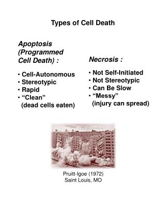
Apoptosis summary • programmed cell death • active process associated with the activation of many genes • requiring energy input • usually involves single cells physiological process of conditioning the proper functioning of the body, both at the stage of embryonic and beyond • The first morphological sign of proving the initiation of cell apoptosis are changes at the nuclear level: • Chromatin undergoes condensation, and is localized just below the cell membrane, • This leads to shrinkage of the whole kernel and its fragmentation and then the entire cell cytoplasm: • This leads to condensation of the cytoplasm • Creation of a specific cell-surface bubbles • The bulges are formed from cell membrane apoptotic bodies, which are structures containing chromatin and cytoplasm, organelles • The final step is the phagocytosis of apoptotic bodies formed
Causes of apoptosis:Physical - ionizing radiation, UV, thermal shockChemicals - cytostatics, free oxygen radicalsBiologicals - glucocorticoids, TNF agent, an anti Fas/APO-1 deficiency, growth substances, absence of cell adhesion, cell organelle damageApoptosis is essential for the proper functioning of the body and occurs continuously under physiological conditions.
Cells undergoing apoptosis in the adult organism:Eye - the eye lens composed of apoptotic cellsIntestine - projections forming cells in the intestinal wall from the base of the villus migrate within a few days of its tip, there will undergo apoptosisSkin - moving toward the deeper layers of the skin and undergo apoptosisThe thymus - T cells matured in the thymus gland, which can not recognize foreign antigens or attack its own tissues, undergo apoptosisUterus - mucosal cells undergo apoptosis and peel off during menstruation
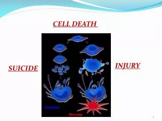
Other types of cell death Cell Autophagy cell digests some elements of its own structure Mitotic catastrophe wrong cytokinesis, no kariokinesis necrosis the nonspecific, passive degradationprocess apoptosis physiological suicideal death
Necrosis - A passive process* (necrosis is a process which can be closely controlled by molecular mechanisms programmed, TNF-α activates the necrosis by forming a complex comprising a protein RIP1 and RIP3) takes place under the influence of all factors physical, chemical and biological. • It is caused by the low and high temperatures, UV light, and bacterial toxins • It refers to a group of cells which swell and lose membrane integrity
Necrosis • Changes in cells that become necrotic include:ATP levels fall, as a result of mitochondrial membrane depolarization, which is a consequence of disturbances in the transport of electrons. • Disturbances in the structure of the cell membrane leads to passive influx of water and ions (mainly calcium and sodium) into the cell, and an increase in their concentration affects the activation of nucleasesrelease of hydrolytic enzymes from lysosomes bursting, which disrupts the structure of other organelles in the cell. • Swollen cell and its organelles disintegrate and the entire contents are released into the intercellular space.Spilled cellular components cause inflammation, which is not in the case of apoptosis