Download
1 / 49
490 likes | 660 Views
Grand Ward Round 17th April 2008. MO presenting: D Ho Consultants-in-charge: Dr V Yong Dr N Tan. Case 1. 39, chinese, male, construction worker Painful right eye with blurring of vision from 1pm on day of presentation. History. While hammering metal, felt speck of metal fly into right eye

E N D
Grand Ward Round17th April 2008 MO presenting: D Ho Consultants-in-charge: Dr V Yong Dr N Tan
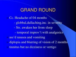
Case 1 • 39, chinese, male, construction worker • Painful right eye with blurring of vision from 1pm on day of presentation
History • While hammering metal, felt speck of metal fly into right eye • At presentation, in right eye • Pain • Redness • Blurring of vision • Not wearing safety goggles at that time
Physical Examination • Visual acuity • Right 6/30, no improvement on pinhole • Left 6/6 • No RAPD
Ocular trauma score • Presenting visual acuity • Presence of endophthalmitis • Globe rupture • Perforating injury • Retinal detachment • Afferent pupillary defect
Left eye infero-nasal corneo-scleral laceration with iris plugging Intra-ocular foreign body Possible lens involvement Provisional Diagnosis
Predictors for poor visual outcome in IOFB • Visual acuity (≥ hand movements) • RAPD • Hyphaema • Vitreous haemorrhage • Uveal prolapse • Retinal detachment • Corneoscleral entrance wound
Management • Decision to proceed with toilet and suture, and examination under anaesthesia • Antibiotic coverage • Recommended • Fluoroquinolones eg moxifloxacin, levofloxacin • Systemic and intra-vitreal • Tetanus immunisation
Intra-operative findings • Corneo-scleral laceration • ? Foreign body at inferior aspect of AC, removed using aspiration cannula
Post-operative findings • Reflectile object on inferior pole of lens
Management • Decision to close up and repeat CT
Repeat CT orbits • Residual intra-ocular foreign body
Management • Decision for repeat re-exploration of globe and removal of IOFB and lens
Management • TPPV • IOFB moved to anterior vitreous with intra-ocular magnet • Pars plana sclerostomy enlarged, FB removed with forceps • Cryo and endolaser
Case 2 • 40, Indian, Male Shipyard worker • Pain over right eye, cheek and forehead
History • Was involved in a sand blast injury at work • Claims to have had blast of sand particles from compressor hit right cheek, eye and forehead • Now presenting with right eye • Blurring of vision • Pain and redness
Physical Examination • Visual acuity • Right eye HM @ 3 ft, no improvement • Left eye 6/12, on PH 6/9 • No RAPD
Physical Examination • Multiple foreign bodies on cornea, conjunctiva, lid and face on right side • Right eye • Diffused sub-conjunctival haemorrhage • AC flare, no cells • Cornea - large epithelial defect • Self sealing scleral laceration at superior temporal region.
Investigation • X-ray: multiple tiny opacities over right orbit, changing in position with look up and look down • CT orbits • Multiple particles embedded in face, conjunctiva and scleral of right globe • Small particels in lens, within AC • No obvious FB in posterior chamber
Management • Decision to preceed with right TPPV, T&S of scleral laceration, exploration, removal of IOFB
Intra-operative findings • Multiple stone foreign bodies embedded in and under conjunctiva superior, nasal, temporal and bulbar areas • Conjunctival laceration at 10 o’clock • Scleral wound at 10 o’clock • Lens intact
Operative procedure • Examination under anaesthesia • Exploration of corneoscleral wounds • Toilet and suture of corneoscleral lacerations • Removal of extraocular foreign bodies
Post-operation • Repeat CT scan done to look for any other IOFB
Investigation • 2 foreign bodies remaining on right globe at 6 and 8 o’clock positions
Post-operation • On review • Patient noted to have FB at 6 o’clock position on gonioscopy • Decision to proceed for repeat removal of FB, with TPPV and internal search
Management • Foreign body in anterior chamber at 6 o’clock removed • Attempted TPPV, but abandoned as unable to create PVD • Thorough internal search for IOFB • None found
Discussion • Delayed vs Early removal • Considerations • Presence of clinical endophthalmitis • Concomitant medical problems of patients • Availability of trained OT personnel
Discussion • Early removal • Within first 24 hours • Advantages • Decrease risk of endophthalmitis • Decrease risk of proliferative vitroretinopathy • Decrease anaesthetic risk
Discussion • Delayed removal • More than 24hours after first operation • Ferrari et al: no difference in functional or anatomical outcome observed with delayed surgery (mean 5.75 days) • Colyer et al: delayed IOFB removal may not be associated with higher risk of endophthalmitis (72hr to 21days)
Discussion • Delayed removal • Advantages • Better integrity of repaired laceration • Less severe anterior segment pathology • Hyphaema resolution • Resorption of corneal oedema • Allow time for PVD to develop • Resorption of some vitreous haemorrhage
References • Yeh S et al. Current trends in the management of intraocular foreign bodies. Curr Opin Ophthalmol 2008 May;19(3):225-33 • Kuhn F et al. A standardized classification of ocular trauma. Opthalmology 1996;103:240-3 • Wickham L et al. Outcomes of surgery for posterior segment intraocular foreign bodies - a retrospective review of 17 years of clinical experience. Graefes Arch Clin Ex Ophthalmol 2006;224:1620-6 • De Souza S et al. Management of posterior segment intraocular foreign bodies: 14 years’ experience. Can J Opthalmol 1999;43:23-9 • Ferrari et al. Early vs late removal of foreign bodies. Retina 2001;21:92-3 • Coyler MH et al. Delayed intraocular foreign body removal in operation iraqi freedom and enduring freedom. Ophthalmology 2007 Aug;114(8):439-47