Download
1 / 67
680 likes | 871 Views
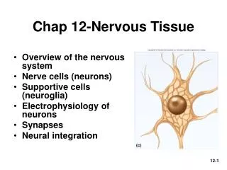
Chapter 12 Nervous Tissue. Controls and integrates all body activities within limits that maintain life Three basic functions sensing changes with sensory receptors fullness of stomach or sun on your face interpreting and remembering those changes reacting to those changes with effectors

E N D
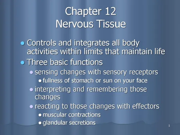
Chapter 12Nervous Tissue • Controls and integrates all body activities within limits that maintain life • Three basic functions • sensing changes with sensory receptors • fullness of stomach or sun on your face • interpreting and remembering those changes • reacting to those changes with effectors • muscular contractions • glandular secretions Tortora & Grabowski 9/e 2000 JWS
Major Structures of the Nervous System • Brain, cranial nerves, spinal cord, spinal nerves, ganglia, enteric plexuses and sensory receptors Tortora & Grabowski 9/e 2000 JWS
Organization of the Nervous System • CNS is brain and spinal cord • PNS is everything else Tortora & Grabowski 9/e 2000 JWS
Nervous System Divisions • Central nervous system (CNS) • consists of the brain and spinal cord • Peripheral nervous system (PNS) • consists of cranial and spinal nerves that contain both sensory and motor fibers • connects CNS to muscles, glands & all sensory receptors Tortora & Grabowski 9/e 2000 JWS
Subdivisions of the PNS • Somatic (voluntary) nervous system (SNS) • neurons from cutaneous and special sensory receptors to the CNS • motor neurons to skeletal muscle tissue • Autonomic (involuntary) nervous systems • sensory neurons from visceral organs to CNS • motor neurons to smooth & cardiacmuscle and glands • sympathetic division (speeds up heart rate) • parasympathetic division (slow down heart rate) • Enteric nervous system (ENS) • involuntary sensory & motor neurons control GI tract • neurons function independently of ANS & CNS Tortora & Grabowski 9/e 2000 JWS
Neurons • Functional unit of nervous system • Have capacity to produce action potentials • electrical excitability • Cell body • single nucleus with prominent nucleolus • Nissl bodies (chromatophilic substance) • rough ER & free ribosomes for protein synthesis • neurofilaments give cell shape and support • microtubules move material inside cell • lipofuscin pigment clumps (harmless aging) • Cell processes = dendrites & axons Tortora & Grabowski 9/e 2000 JWS
Parts of a Neuron Neuroglial cells Nucleus with Nucleolus Axons or Dendrites Cell body Tortora & Grabowski 9/e 2000 JWS
Dendrites • Conducts impulses towards the cell body • Typically short, highly branched & unmyelinated • Surfaces specialized for contact with other neurons • Contains neurofibrils & Nissl bodies Tortora & Grabowski 9/e 2000 JWS
Axons • Conduct impulses away from cell body • Long, thin cylindrical process of cell • Arises at axon hillock • Impulses arise from initial segment (trigger zone) • Side branches (collaterals) end in fine processes called axon terminals • Swollen tips called synaptic end bulbs contain vesicles filled with neurotransmitters Tortora & Grabowski 9/e 2000 JWS Synaptic boutons
Axonal Transport • Cell body is location for most protein synthesis • neurotransmitters & repair proteins • Axonal transport system moves substances • slow axonal flow • movement in one direction only -- away from cell body • movement at 1-5 mm per day • fast axonal flow • moves organelles & materials along surface of microtubules • at 200-400 mm per day • transports in either direction • for use or for recycling in cell body Tortora & Grabowski 9/e 2000 JWS
Axonal Transport & Disease • Fast axonal transport route by which toxins or pathogens reach neuron cell bodies • tetanus (Clostridium tetani bacteria) • disrupts motor neurons causing painful muscle spasms • Bacteria enter the body through a laceration or puncture injury • more serious if wound is in head or neck because of shorter transit time Tortora & Grabowski 9/e 2000 JWS
Functional Classification of Neurons • Sensory (afferent) neurons • transport sensory information from skin, muscles, joints, sense organs & viscera to CNS • Motor (efferent) neurons • send motor nerve impulses to muscles & glands • Interneurons (association) neurons • connect sensory to motor neurons • 90% of neurons in the body Tortora & Grabowski 9/e 2000 JWS
Structural Classification of Neurons • Based on number of processes found on cell body • multipolar = several dendrites & one axon • most common cell type • bipolar neurons = one main dendrite & one axon • found in retina, inner ear & olfactory • unipolar neurons = one process only(develops from a bipolar) • are always sensory neurons Tortora & Grabowski 9/e 2000 JWS
Association or Interneurons • Named for histologist that first described them or their appearance Tortora & Grabowski 9/e 2000 JWS
Neuroglial Cells • Half of the volume of the CNS • Smaller cells than neurons • 50X more numerous • Cells can divide • rapid mitosis in tumor formation (gliomas) • 4 cell types in CNS • astrocytes, oligodendrocytes, microglia & ependymal • 2 cell types in PNS • schwann and satellite cells Tortora & Grabowski 9/e 2000 JWS
Astrocytes • Star-shaped cells • Form blood-brain barrier by covering blood capillaries • Metabolize neurotransmitters • Regulate K+ balance • Provide structural support Tortora & Grabowski 9/e 2000 JWS
Oligodendrocytes • Most common glial cell type • Each forms myelin sheath around more than one axons in CNS • Analogous to Schwann cells of PNS Tortora & Grabowski 9/e 2000 JWS
Microglia • Small cells found near blood vessels • Phagocytic role -- clear away dead cells • Derived from cells that also gave rise to macrophages & monocytes Tortora & Grabowski 9/e 2000 JWS
Ependymal cells • Form epithelial membrane lining cerebral cavities & central canal • Produce cerebrospinal fluid (CSF) Tortora & Grabowski 9/e 2000 JWS
Satellite Cells • Flat cells surrounding neuronal cell bodies in peripheral ganglia • Support neurons in the PNS ganglia Tortora & Grabowski 9/e 2000 JWS
Schwann Cell • Cells encircling PNS axons • Each cell produces part of the myelin sheath surrounding an axon in the PNS Tortora & Grabowski 9/e 2000 JWS
Axon Coverings in PNS • All axons surrounded by a lipid & protein covering (myelin sheath) produced by Schwann cells • Neurilemma is cytoplasm & nucleusof Schwann cell • gaps called nodes of Ranvier • Myelinated fibers appear white • jelly-roll like wrappings made of lipoprotein = myelin • acts as electrical insulator • speeds conduction of nerve impulses • Unmyelinated fibers • slow, small diameter fibers • only surrounded by neurilemma but no myelin sheath wrapping Tortora & Grabowski 9/e 2000 JWS
Myelination in PNS • Schwann cells myelinate (wrap around) axons in the PNS during fetal development • Schwann cell cytoplasm & nucleus forms outermost layer of neurolemma with inner portion being the myelin sheath • Tube guides growing axons that are repairing themselves Tortora & Grabowski 9/e 2000 JWS
Myelination in the CNS • Oligodendrocytes myelinate axons in the CNS • Broad, flat cell processes wrap about CNS axons, but the cell bodies do not surround the axons • No neurilemma is formed • Little regrowth after injury is possible due to the lack of a distinct tube or neurilemma Tortora & Grabowski 9/e 2000 JWS
Gray and White Matter • White matter = myelinated processes (white in color) • Gray matter = nerve cell bodies, dendrites, axon terminals, bundles of unmyelinated axons and neuroglia (gray color) • In the spinal cord = gray matter forms an H-shaped inner core surrounded by white matter • In the brain = a thin outer shell of gray matter covers the surface & is found in clusters called nuclei inside the CNS Tortora & Grabowski 9/e 2000 JWS
Electrical Signals in Neurons • Neurons are electrically excitable due to the voltage difference across their membrane • Communicate with 2 types of electric signals • action potentials that can travel long distances • graded potentials that are local membrane changes only • In living cells, a flow of ions occurs through ion channels in the cell membrane Tortora & Grabowski 9/e 2000 JWS
Two Types of Ion Channels • Leakage (nongated) channels are always open • nerve cells have more K+ than Na+ leakage channels • as a result, membrane permeability to K+ is higher • explains resting membrane potential of -70mV in nerve tissue • Gated channels open and close in response to a stimulus results in neuron excitability • voltage-gated open in response to change in voltage • ligand-gated open & close in response to particular chemical stimuli (hormone, neurotransmitter, ion) • mechanically-gated open with mechanical stimulation Tortora & Grabowski 9/e 2000 JWS
Gated Ion Channels Tortora & Grabowski 9/e 2000 JWS
Resting Membrane Potential • Negative ions along inside of cell membrane & positive ions along outside • potential energy difference at rest is -70 mV • cell is “polarized” • Resting potential exists because • concentration of ions different inside & outside • extracellular fluid rich in Na+ and Cl • cytosol full of K+, organic phosphate & amino acids • membrane permeability differs for Na+ and K+ • 50-100 greater permeability for K+ • inward flow of Na+ can’t keep up with outward flow of K+ • Na+/K+ pump removes Na+ as fast as it leaks in Tortora & Grabowski 9/e 2000 JWS
Graded Potentials • Small deviations from resting potential of -70mV • hyperpolarization = membrane has become more negative • depolarization = membrane has become more positive Tortora & Grabowski 9/e 2000 JWS
How do Graded Potentials Arise? • Source of stimuli • mechanical stimulation of membranes with mechanical gated ion channels (pressure) • chemical stimulation of membranes with ligand gated ion channels (neurotransmitter) • Graded/postsynaptic/receptor or generator potential • ions flow through ion channels and change membrane potential locally • amount of change varies with strength of stimuli • Flow of current (ions) is local change only Tortora & Grabowski 9/e 2000 JWS
Action Potential • Series of rapidly occurring events that change and then restore the membrane potential of a cell to its resting state • Ion channels open, Na+ rushes in (depolarization), K+ rushes out (repolarization) • All-or-none principal = with stimulation, either happens one specific way or not at all (lasts 1/1000 of a second) • Travels (spreads) over surface of cell without dying out Tortora & Grabowski 9/e 2000 JWS
Depolarizing Phase of Action Potential • Chemical or mechanical stimuluscaused a graded potential to reachat least (-55mV or threshold) • Voltage-gated Na+ channels open& Na+ rushes into cell • in resting membrane, inactivation gate of sodium channel is open & activation gate is closed (Na+ can not get in) • when threshold (-55mV) is reached, both open & Na+ enters • inactivation gate closes again in few ten-thousandths of second • only a total of 20,000 Na+ actually enter the cell, but they change the membrane potential considerably(up to +30mV) • Positive feedback process Tortora & Grabowski 9/e 2000 JWS
Repolarizing Phase of Action Potential • When threshold potential of-55mV is reached, voltage-gated K+ channels open • K+ channel opening is muchslower than Na+ channelopening which caused depolarization • When K+ channels finally do open, the Na+ channels have already closed (Na+ inflow stops) • K+ outflow returns membrane potential to -70mV • If enough K+ leaves the cell, it will reach a -90mV membrane potential and enter the after-hyperpolarizing phase • K+ channels close and the membrane potential returns to the resting potential of -70mV Tortora & Grabowski 9/e 2000 JWS
Refractory Period of Action Potential • Period of time during whichneuron can not generateanother action potential • Absolute refractory period • even very strong stimulus willnot begin another AP • inactivated Na+ channels must return to the resting state before they can be reopened • large fibers have absolute refractory period of 0.4 msec and up to 1000 impulses per second are possible • Relative refractory period • a suprathreshold stimulus will be able to start an AP • K+ channels are still open, but Na+ channels have closed Tortora & Grabowski 9/e 2000 JWS
The Action Potential: Summarized • Resting membrane potential is -70mV • Depolarization is the change from -70mV to +30 mV • Repolarization is the reversal from +30 mV back to -70 mV) Tortora & Grabowski 9/e 2000 JWS
Propagation of Action Potential • An action potential spreads (propagates) over the surface of the axon membrane • as Na+ flows into the cell during depolarization, the voltage of adjacent areas is effected and their voltage-gated Na+ channels open • self-propagating along the membrane • The traveling action potential is called a nerve impulse Tortora & Grabowski 9/e 2000 JWS
Local Anesthetics • Prevent opening of voltage-gated Na+ channels • Nerve impulses cannot pass the anesthetized region • Novocaine and lidocaine Tortora & Grabowski 9/e 2000 JWS
Continuous versus Saltatory Conduction • Continuous conduction (unmyelinated fibers) • step-by-step depolarization of each portion of the length of the axolemma • Saltatory conduction • depolarization only at nodes of Ranvier where there is a high density of voltage-gated ion channels • current carried by ions flows through extracellular fluid from node to node Tortora & Grabowski 9/e 2000 JWS
Saltatory Conduction • Nerve impulse conduction in which the impulse jumps from node to node Tortora & Grabowski 9/e 2000 JWS
Speed of Impulse Propagation • The propagation speed of a nerve impulse is not related to stimulus strength. • larger, myelinated fibers conduct impulses faster due to size & saltatory conduction • Fiber types • A fibers largest (5-20 microns & 130 m/sec) • myelinated somatic sensory & motor to skeletal muscle • B fibers medium (2-3 microns & 15 m/sec) • myelinated visceral sensory & autonomic preganglionic • C fibers smallest (.5-1.5 microns & 2 m/sec) • unmyelinated sensory & autonomic motor Tortora & Grabowski 9/e 2000 JWS
Encoding of Stimulus Intensity • How do we differentiate a light touch from a firmer touch? • frequency of impulses • firm pressure generates impulses at a higher frequency • number of sensory neurons activated • firm pressure stimulates more neurons than does a light touch Tortora & Grabowski 9/e 2000 JWS
Action Potentials in Nerve and Muscle • Entire muscle cell membrane versus only the axon of the neuron is involved • Resting membrane potential • nerve is -70mV • skeletal & cardiac muscle is closer to -90mV • Duration • nerve impulse is 1/2 to 2 msec • muscle action potential lasts 1-5 msec for skeletal & 10-300msec for cardiac & smooth • Fastest nerve conduction velocity is 18 times faster than velocity over skeletal muscle fiber Tortora & Grabowski 9/e 2000 JWS
Comparison of Graded & Action Potentials • Origin • GPs arise on dendrites and cell bodies • APs arise only at trigger zone on axon hillock • Types of Channels • AP is produced by voltage-gated ion channels • GP is produced by ligand or mechanically-gated channels • Conduction • GPs are localized (not propagated) • APs conduct over the surface of the axon Tortora & Grabowski 9/e 2000 JWS
Comparison of Graded & Action Potentials • Amplitude • amplitude of the AP is constant (all-or-none) • graded potentials vary depending upon stimulus • Duration • The duration of the GP is as long as the stimulus lasts • Refractory period • The AP has a refractory period due to the nature of the voltage-gated channels, and the GP has none. Tortora & Grabowski 9/e 2000 JWS
Signal Transmission at Synapses • 2 Types of synapses • electrical • ionic current spreads to next cell through gap junctions • faster, two-way transmission & capable of synchronizing groups of neurons • chemical • one-way information transfer from a presynaptic neuron to a postsynaptic neuron • axodendritic -- from axon to dendrite • axosomatic -- from axon to cell body • axoaxonic -- from axon to axon Tortora & Grabowski 9/e 2000 JWS
Chemical Synapses • Action potential reaches end bulb and voltage-gated Ca+ 2 channels open • Ca+2 flows inward triggering release of neurotransmitter • Neurotransmitter crosses synaptic cleft & binding to ligand-gated receptors • the more neurotransmitter released the greater the change in potential of the postsynaptic cell • Synaptic delay is 0.5 msec • One-way information transfer Tortora & Grabowski 9/e 2000 JWS
Excitatory & Inhibitory Potentials • The effect of a neurotransmitter can be either excitatory or inhibitory • a depolarizing postsynaptic potential is called an EPSP • it results from the opening of ligand-gated Na+ channels • the postsynaptic cell is more likely to reach threshold • an inhibitory postsynaptic potential is called an IPSP • it results from the opening of ligand-gated Cl- or K+ channels • it causes the postsynaptic cell to become more negative or hyperpolarized • the postsynaptic cell is less likely to reach threshold Tortora & Grabowski 9/e 2000 JWS
Removal of Neurotransmitter • Diffusion • move down concentration gradient • Enzymatic degradation • acetylcholinesterase • Uptake by neurons or glia cells • neurotransmitter transporters • Prozac = serotonin reuptake inhibitor Tortora & Grabowski 9/e 2000 JWS
Spatial Summation • Summation of effects of neurotransmitters released from several end bulbs onto one neuron Tortora & Grabowski 9/e 2000 JWS