Download
1 / 89
890 likes | 1.1k Views
ULCERATIVE, VESICULAR, AND BULLOUS LESIONS (lecture 2). Dr rami aljuaidi. introduction. Many diseases have semilar apperances Lesions have nonspecific appearance on the oral mucosa Diagnosis require:

E N D
ULCERATIVE, VESICULAR, ANDBULLOUS LESIONS (lecture 2) Dr rami aljuaidi
introduction • Many diseases have semilar apperances • Lesions have nonspecific appearance on the oral mucosa • Diagnosis require: • 1-detailed history based on review of systemes and associated symptomes 2- clinical examination which require knowledge of dermatology especially the elementary lesions
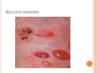
1. Macules. Well-circumscribed, flat lesions that are noticeable because of their change from normal skin color . 2. Papules. Solid lesions raised above the skin surface that are smaller than 1 cm in diameter 3. Plaques. Solid raised lesions that are over 1 cm in diameter; they are large papules. 4. Nodules. These lesions are present deep in the dermis, and the epidermis can be easily moved over them. 5. Vesicles. Elevated blisters containing clear fluid that are under 1 cm in diameter. 6. Bullae. Elevated blisterlike lesions containing clear fluid that are over 1 cm in diameter. 7. Erosions.Moist red lesions often caused by the rupture of vesicles or bullae as well as trauma. 8. Pustules. Raised lesions containing purulent material. 9. Ulcers. A defect in the epithelium; it is a well-circumscribed depressed lesion over which the epidermal layer has been lost. 10. Purpura. Reddish to purple flat lesions caused by blood from vessels leaking into the subcutaneous tissue. Classified by size as petechiae or ecchymoses,
history • 3 pieces of informations must be obtained to categorize the disease and to simplify the diagnosis: • 1-acute or chronic • 2-primary or recurrent • 3-sigle or multiples
THE PATIENT WITH ACUTE MULTIPLELESIONS • ▼ • Herpesvirus Infections • Primary Herpes Simplex Virus Infections • Coxsackievirus Infections • Varicella-Zoster Virus Infection • Erythema Multiforme • Contact Allergic Stomatitis • Oral Ulcers Secondary to Cancer Chemotherapy • Acute Necrotizing Ulcerative Gingivitis
▼ THE PATIENT WITH RECURRING ORALULCERS • Recurrent Aphthous Stomatitis • Behçet’s Syndrome • Recurrent Herpes Simplex Virus Infection
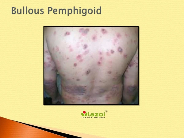
▼ THE PATIENT WITH CHRONIC MULTIPLELESIONS • Pemphigus • Subepithelial Bullous Dermatoses • Herpes Simplex Virus Infection in Immunosuppressed Patients
▼ THE PATIENT WITH SINGLE ULCERS • Traumatic ulcer • Malignant ulcer • Syphilis(primary and tertiary) • Histoplasmosis • Blastomycosis • Mucormycosis
Acute multiple lesionsherpesvirus infections • 8 ef them are known to cause humain infections: Hsv1,Hsv2( oral mucosal diseases),varicella-zoster virus, cytomegalovirus (salivary glands diseases in immunosupressed patients)),EBV,Hhv6 (roseola infantum,mononucleoses,pneumonitis and bone marow suppression), Hhv7( unspecific),Hhv8( kaposi sarcoma,lymphoma)
HSV1 causes a majority of cases of oral and pharyngeal infection,meningoencephalitis, and dermatitis above the waist; HSV2 is implicated in most genital infections. Humans are the only natural reservoir of HSV infection, and spread occurs by direct intimate contact with lesions or secretions from an asymptomatic carrier Latency, a characteristic of all herpesviruses, peripheral tissue injury from trauma or sunburn, fever, or immunosuppression can cause reactivation of the virus. recent evidence has demonstrated that reactivation of HSV is the most common cause of bell’s pulsy. an increased incidence of HSV2 serum antibodies or positive HSV2 cultures in patients with cervical carcinoma. .
Primary Herpes Simplex Virus Infections • There are approximately 600,000 new cases of primary HSV infections per year in the United States. Primary HSV infection occurs in patients who do not have immunity resulting from previous contact with the virus. HSV is contracted after intimate • contact with an individual who has active HSV primary • or recurrent lesions. Primary HSV may also be spread by asymptomatic shedders with HSV present in salivary secretions. • The majority of oral HSV infections is caused by HSV1, • but primary oral HSV2 infections may also occur • Infection of the fingers (herpetic whitlows) of health professionals may occur during treatment • of infected patients
Primary HSV infection of the newborn was previously believed to be caused by direct contact with vaginal HSV lesions during birth, but it has now been established that a majority of mothers giving birth to children with primary HSV are asymptomatic carriers without lesions. These infections of the newborn result in viremia and disseminated infection of the brain, liver, adrenals, and lungs.
Newborns of mothers with antibody titers are protected by placentally transferred antibodies during the first 6 months of life. After 6 months of age, the incidence of primary HSV1 infection increases. The incidence of primary HSV1 infection reaches a peak between 2 and 3 years of age. Incidence of primary HSV2 infection does not increase until the age when sexual activity begins The incidence of primary herpes infection has been shown to vary according to socioeconomic group.
CLINICAL MANIFESTATIONS OF PRIMARY ORAL HERPES • The • incubation period is most commonly 5 to 7 days but may • range from 2 to 12 days. • Patients with primary oral herpes have a history of generalized • prodromal symptoms that precede the local lesions by • 1 or 2 days. This information is helpful in differentiating this • viral infection from allergic stomatitis or erythema multiforme, • in which local lesions and systemic symptoms appear • together.These generalized symptoms include fever, headache, • malaise, nausea, and vomiting. A negative past history of recurrent • herpes labialis and a positive history of direct intimate • contact with a patient with primary or recurrent herpes are • also helpful in making the diagnosis.
Approximately 1 or 2 days after the prodromal symptoms occur, small vesicles appear on the oral mucosa; these are thin-walled vesicles surrounded by an inflammatory base The vesicles quickly rupture, leaving shallow round discrete ulcers. The lesions occur on all portions of the mucosa. As the disease progresses, several lesions may coalesce, forming larger irregular lesions.
Acute marginal gingivitis characteristic of primary HSV infection. A, mandibular anterior gingiva; B, vesicles and inflammation around mandibular molars.
An important diagnostic criterion in this disease is the appearance of generalized acute marginal gingivitis. The entire gingiva is edematous
Several small gingival ulcers are often present. Examination of the posterior pharynx reveals inflammation, and the submandibular and cervical lymph nodes are characteristically enlarged and tender. On occasion, primary HSV may cause lesions of the labial and facial skin without intraoral lesions. Primary HSV in otherwise healthy children is a self-limiting disease. The fever ordinarily disappears within 3 or 4 days, and the lesions begin healing in a week to 10 days, although HSV may continue to be present in the saliva for up to a month after the onset of disease.
LABORATORY DIAGNOSIS • In some patients, especially adults, may have a less typical clinical picture,making the diagnosis more difficult. This is especially important when distinguishing primary herpes from erythema multiforme since proper therapy differs significantly. • The following laboratory tests are helpful in the diagnosis of a primary herpes infection. • Cytology ballooning degeneration and multinucleated giant cells • HSV Isolation • . Antibody Titers • .
Cytology smear stained with Giemsa, demonstrating multinucleated giant cells.
TREATMENT • A significant advance in the management of herpes simplex • infections was the discovery of acyclovir, which has no effect • on normal cells but inhibits DNA replication in HSV-infected • cells. Acyclovir has been shown to be effective in the treatment • of primary oral HSV in children when therapy was • started in the first 72 hours.
Coxsackievirus Infections • Coxsackieviruses are ribonucleic acid (RNA) enteroviruses, separated into two groups, A and B. There are 24 known types of coxsackievirus group A and 6 types of coxsackievirus group B. • These viruses cause hepatitis, meningitis, myocarditis, pericarditis, and acute respiratory disease. • Three clinical types of infection of the oral region that have been described are usually caused by group A coxsackieviruses: herpangina, hand foot-and-mouth disease, and acute lymphonodular pharyngitis. • Types of coxsackievirus A have also been described as • causing a rare mumpslike form of parotitis.
HERPANGINA • Coxsackievirus A4 has been shown to cause a majority of cases, herpangina may be seen more than once in the same patient. • Unlike herpes simplex infections,which occur at a constant rate,herpangina frequently occurs in epidemics that have their highest incidence from June to October. The majority of cases affect young children ages 3 through 10, but infection of adolescents and adults is not uncommon
Clinical Manifestations. After a 2- to 10-day incubation period, the infection begins with generalized symptoms of fever, chills, and anorexia. The fever and other symptoms are generally milder than those experienced with primary HSV infection. The patient complains of sore throat, dysphagia, and occasionally sore mouth. Lesions start as punctate macules, which quickly evolve into papules and vesicles involving the posterior pharynx, tonsils, and soft palate. Lesions are found less frequently on the buccal mucosa, tongue, and hard palate. Within 24 to 48 hours, the vesicles rupture, forming small 1 to 2 mm ulcers. The disease is usually mild and heals without treatment in 1 week.
Herpangina may be clinically distinguished from primary HSV infection by several criteria: 1. Herpangina occurs in epidemics; HSV infections do not. 2. Herpangina tends to be milder than HSV infection. 3. Lesions of herpangina occur on the pharynx and posterior portions of the oral mucosa, whereas HSV primarily affects the anterior portion of the mouth. 4. Herpangina does not cause a generalized acute gingivitis like that associated with primary HSV infection. 5. Lesions of herpangina tend to be smaller than those of HSV.
Laboratory Studies. • A smear taken from the base of a fresh • vesicle and stained with Giemsa will not show ballooning degeneration or multinucleated giant cells. This helps to distinguish herpangina from herpes simplex and herpes zoster, • which do show these changes. • Treatment. Herpangina is a self-limiting disease, and treatment is supportive • ,
ACUTE LYMPHONODULAR PHARYNGITIS • This is a variant of herpangina caused by coxsackievirus A10. • The distribution of the lesions is the same as in herpangina,but yellow-white nodules appear that do not progress to vesicles • or ulcers. The disease is self-limiting, and only supportive care is indicated.
HAND-FOOT-AND-MOUTH DISEASE • Hand-foot-and-mouth disease is caused by infection with coxsackievirus A16 in a majority of cases, • The disease is characterized by low-grade fever, oral vesicles and ulcers, and nonpruritic macules, papules, and vesicles, particularly on the extensor surfaces of the hands and feet. The oral lesions are more extensive than are those described for herpangina, and lesions of the hard palate, tongue, and buccal mucosa are common. • Severe cases with central nervous system involvement,myocarditis, and pulmonary edema have been reported in epidemics
The patients ranged in age from 8 months to 33 years,with 75% of cases occurring below 4 years of age. The clinical manifestations lasted 3 to 7 days. The most common complaint of patients was a sore mouth, and, clinically, lesions involving the oral mucosa. Because of the frequent oral involvement, dentists are more likely to see patients with this disease than with herpangina, and they should remember to examine the hands and feet for maculopapular and vesicular lesions when patients present with an acute stomatitis and fever. Treatment is supportive.
Varicella-Zoster Virus Infection • Varicella zoster (VZV) is a herpesvirus,. responsible for two major clinical • infections of humans: chickenpox(varicella) and shingles (herpes zoster [HZ]). • Chickenpox is a generalized primary infection that occurs the first time an • individual contacts the virus. This is analogous to the acute herpetic • gingivostomatitis of herpes simplex virus. • After the primary disease is healed,VZV becomes latent in the dorsal root • ganglia of spinal nerves or ganglia of cranial nerves. A child • without prior contact with VZV can develop chickenpox after contact with an • individual with HZ. • In 3 to 5 of every 1,000 individuals, VZV becomes reactivated, causing lesions • of localized herpes zoster.
The incidence of HZ increases with age or immunosuppression. Patients who are immunocompromised have an increased susceptibility to severe and potentially fatal HZ. These HZ infections may be deep-seated and disseminated, causing pneumonia, meningoencephalitis, and hepatitis; however, otherwise normal patients who develop HZ do not have a significant incidence of underlying malignancy.
General Findings. Chickenpox is a childhood disease characterized by mild systemic symptoms and a generalized intensely pruritic eruption of maculopapular lesions that rapidly develop into vesicles on an erythematous base. Oral vesicles that rapidly change to ulcers may be seen, but the oral lesions are not an important symptomatic, diagnostic, or management problem.
HZ commonly has a prodromal period of 2 to 4 days,when shooting pain, paresthesia, burning, and tenderness appear along the course of the affected nerve.Unilateral vesicles on an erythematous base then appear in clusters, chiefly along the course of the nerve, giving the characteristic clinical picture of single dermatome involvement. Some lesions spread by viremia occur outside the dermatome. The vesicles turn to scabs in 1 week, and healing takes place in 2 to 3 weeks.
The Nerve most commonly affected with HZ : the first division of the trigeminal nerve When the full clinical picture of HZ is present with pain and unilateral vesicles, the diagnosis is not difficult. Diagnostic problems arise during the prodromal period, when pain is present without lesions. Unnecessary surgery has been performed because of the diagnosis of acute appendicitis, cholecystitis, or dental pulpitis. A more difficult diagnostic problem is pain caused by VZ virus without lesions developing along the course of the nerve (zoster sine herpete; zoster sine eruptione). Diagnosis in these cases is based on clinical symptoms and serologic evidence of a rising antibody titer.
HZ may also occasionally affect motor nerves. HZ of the sacral region may cause paralysis of the bladder. The extremities and diaphragm have also been paralyzed during episodes of HZ
The most common complication of HZ is postherpetic neuralgia, which is defined as pain remaining for over a month after the mucocutaneous lesions have healed, although some clinicians do not use the term postherpetic neuralgia unless the pain has lasted for at least 3 months after the healing of the lesions. The overall incidence of postherpetic neuralgia is 12 to 14%, but the risk increases significantly after the age of 60 years, most likely due to the decline in cell-mediated immunity.
Oral Findings • . Herpes zoster involves one of the divisions of the trigeminal nerve in 18 to 20% of cases, but the ophthalmic branch is affected several times more frequently than are the second or third divisions. HZ of the first division can lead to blindness • Facial and intraoral lesions are characteristic of HZ involving the second and third divisions of the trigeminal nerve.
Each individual oral lesion of HZ resembles lesions seen in herpes simplex infections. The diagnosis is based on a history of pain and the unilateral nature and segmental distribution of the lesions .When the clinical appearance is typical and vesicles are present, oral HZ can be distinguished clinically from other acute multiple lesions of the mouth,which are bilateral and are not preceded or accompanied by pain along the course of one trigeminal nerve
HZ has been associated with dental anomalies and severe scarring of the facial skin when trigeminal HZ occurs during tooth formation. Pulpal necrosis and internal root resorption have also been related to HZ. In immunocompromised patients, large chronic HZ lesions have been described that have led to necrosis of underlying bone and exfoliation of teeth.
HZ of the geniculate ganglion, Ramsay Hunt syndrome, is a rare form of the disease characterized by Bell’s palsy, unilateral vesicles of the external ear, and vesicles of the oral mucosa.
isolated oral HZ can be misdiagnosed, particularly when erythema, edema, and nonspecific ulceration are seen without the presence of intact vesicles and when prodromal pain is present prior to the appearance of the characteristic lesions and in zoster sine eruptione. In these cases, a cytology smear or viral culture is often necessary for diagnosis.
LABORATORY FINDINGS • Cytology is a rapid method of evaluation that can be used in • cases in which the diagnosis is uncertain. • Fluorescent-antibody conjugated monoclonal antibodies • viral isolation • Demonstration of a rising antibody titer
TREATMENT • Management should be directed toward shortening the course of the disease, preventing postherpetic neuralgia in patients over 50 years of age, and preventing dissemination in • immunocompromised patients. Acyclovir or the newer antiherpes drugs valacyclovir or famciclovir accelerate healing and reduce acute pain, but they do not reduce the incidence of postherpetic neuralgia. The newer drugs have greater bioavailability and are more effective in the treatment of HZ. • The use of systemic corticosteroids to prevent postherpetic neuralgia in patients over 50 years of age is controversial;
Erythema Multiforme • Erythema multiforme (EM) is an acute inflammatory disease • of the skin and mucous membranes that causes a variety of • skin lesions—hence the name “multiforme.” The oral lesions, • typically inflammation accompanied by rapidly rupturing • vesicles and bullae, are often an important component of the • clinical picture and are occasionally the only component. • Erythema multiforme may occur once or recur, and it should • be considered in the diagnosis of multiple acute oral ulcers • . There is also a rare chronic form of EM. EM has several clinical presentations: • a milder self-limiting form and severe life-threatening • forms that may present as either Stevens-Johnson syndrome or • toxic epidermal necrolysis (TEN).
ETIOLOGY • EM is an immune-mediated disease that may be initiated either by deposition of immune complexes in the superficial microvasculature of skin and mucosa, or cell-mediated immunity.
The most common triggers for episodes of EM are herpes simplex virus and drug reactions. The drugs most frequently associated with EM reactions are oxycam nonsteroidal antiinflammatory drugs (NSAIDs); sulfonamides; anticonvulsants such as carbamazepine, phenobarbital, and phenytoin; trimethoprim-sulfonamide combinations, allopurinol, and penicillin. A majority of the severe cases of Stevens-Johnson syndrome or TEN are caused by drug reactions.
The relationship of HSV to episodes of EM has been known for over 50 years, but improved diagnostic techniques, have demonstrated that herpes-associated EM is a common form of the disease, accounting for at least 20 to 40% of the cases of single episodes of EM and approximately 80% of recurrent EM. Herpes antigens have been demonstrated in the skin and immunocomplexes obtained from patients with EM. Many investigators now believe that the major cause of EM is a cellular immune response to HSV antigens deposited in keratinocytes of the skin and mucosa.. Oral mucosal lesions were detected in 8 of 12 children with HSV-associated EM