Download
1 / 30
310 likes | 488 Views
Inflammation. MD , Professor BONDARENKO Yu.I. Find some one common in all words !!!. Appendic it is Artr it is Cholecyst it is Hepat it is Gastr it is Myocard it is Stomat it is Peridont it is Pulp it is. INFLAMMATION ( definition of the notion ).

E N D
Inflammation MD, Professor BONDARENKO Yu.I.
Find some one common in all words!!! Appendicitis Artritis Cholecystitis Hepatitis Gastritis Myocarditis Stomatitis Peridontitis Pulpitis
INFLAMMATION(definition of the notion) It is atypical pathological process, which arises after damage of tissue and consists of three main vessel-tissue components • alteration; • violation of microcirculation, exudation and migration of leucocytes; • proliferation. _______________________________________ Inflammation, as a typical pathological process has common regularities, which always are present and don’t depend on the cause, localization, species of an organism and it’s individual features
CLASSIFICATION Depending on clinical course Depending on manifestation of clinical signs normoergic aqute chronic hypoergic hyperergic Depending on any stage dominance Exudative Alterative proliferative
Etiology Force and duration of any agent influence should be stronger, than adaptive possibilities of a tissue • Endogen • - Cholic acids • Complex antigen-antibody • Hematoma • Product of intoxication Exogen Physical(alien bodies, hard pressure on a tissue, high and low temperature, ionizing and ultra-violet rays, high and low barometric pressure, electrical current) Chemical(acids, alkalies, salts of heavy metals) Biological (microorganisms - bacterias, viruses, mycotic agents; animal organisms - worms, insects).
STAGES 1 – alteration 2 – exudation 3 - proliferation alteration exudation proliferation
LOCAL SIGNS (by Cels and Gallen) REDNESS (lat. -rubor) -is the result of arterial hyperemia SWELLING (lat. - tumor) – is the result of exudation HEAT (lat. -calor) - is the result of arterial hyperemia and impermanent intensification of metabolism in the focus of inflammation. PAIN (lat. -dolor)- pain is the result of the painful receptors irritation by biological active substances, metabolites, and pressingof painful receptors by exudate LOSS of a FUNCTION (lat. -functio laesa)is the result of the functional active tissue injury. Roman physician Celsus described four signs(swelling, redness, heat, and pain. Greek physician Galen added fifth sing - loss of the function.
GENERALSIGNS LEUCOCYTOSIS – is the result of leucocytes outcome from depot, leucocytes proliferation FIVER – results from IL-1 influence on thermoregulative centre (excreted by macrophages and neutrophyles) PROTEINS OF AQUTE FASE of inflammation – its content increases in the blood on 50 %, they are synthesized mainly in liver, play protective role (inhibitors of proteinases – antitripsine; antioxidants – haptoglobin, ceruloplasmin; IgG, С-reactive proteinе) ESR increase – inflammation couses accumulation of big mass proteins in the blood (globulines, fibrinigen), they adsorb on erythrocytes, decreas surface negative charge and conduceerythrocytesaggregation INTOXICATION– is the result of necrotical substances income in the blood fron area of inflammation
Pathogenesis • Primary alterationis the result of pathological agent influence on a tissue • Secondary alterationis the consequence of the primary alteration and that arises even at the absence of the damaging agent. Metabolism disorder (local acidosis, hyperosmia,hyperoncia), violation of microcirculation, free radicals formation, biological active substances action, lysosomal enzymes (damaged cells origin) conduce its development Damage of the tissue and formation of the biological active substances are the main effects of the alteration
Mediators of the inflammation It is the biological active substances, whichsynthesized or excreted in area of the inflammation and conduce its proression CELLULAR histamine serotonin lymphokines (IL-1, IL-6 et all.) monokines prostaglandines leucotriens lysosomal enzymes HUMORAL complement system proteins bradykinin kallidin Coagulative proteins
HISTAMINE vasodilation increases permeability of the capillaries activation of leucocytes emigration Stimulation of phagocytosis increases adhesive property of the vessels endothelium PAIN
INTERLEUKINE-1 Muscles ----------pain Joins ------------ pain CNS -------------- somnolence Liver ----------------- Protein synthesis activation Thermoregulative center ---------------- fiver
MICROCIRCULATION VIOLATION І stage SHORT-TIME SPASM OF THE VESSELS lasts from 10-20 seconds up to several minutes Y. Kongame has discovered that in experiment on frogs ІІ stage ARTERIAL HYPEREMIA duration - 20-30 minutes, but not more than 1 hour ІІІ stage VENOUS HYPEREMIA ІVstage PRESTASIS Yung Kongame 1839-1884 Vstage STASIS
EXUDATION Blood plasma penetration through the vessels wall and accumulation in the area of the injured tissue. It conducesswelling, painand loss of function Exudation deepensmicrocirculationviolationbecause haemoconcentration (increased viscosity of the blood), aggregationof the erythrocytes and platelets. But in this time vital conditions for microorganisms became worse.
MECHANISM OF EXUDATION • Increasing of vesselpermeability • Increasing of hydrostatic pressurein the vessels • Increasing of oncotic and osmotic pressure in an inflammatory area Vascular wall Blood plassma cells
Migration of the leucocytes Stages - edge standing - penetration through the vessels wall - move in area of inflammation In most cases of acute inflammation neutrophyles migrate the first (that process lasts 6-24 hours). In 24-48 hours monocytes emigrate most actively. Lymphocytes emigrate a little bit later.
Phagocytosis Stagses Approchment Attachment Ingestion digestion
Cells of inflammation Neutrophil Lymphocyte Tissuel macrophages
Types of exudates Serous Fibrinous Purulent Rotten Hemorrhagic
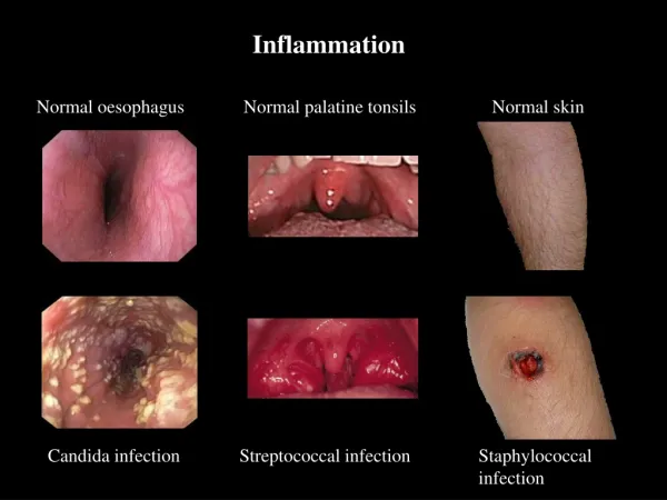
INFLAMMATION TYPES ACCORDING TO EXUDATEKIND SEROUS INFLAMMATION develops in mucous and serous membranes, interstitial tissue, skin, and kidneys glomes capsules. The amount of cells in the serous exudate is not large. The serous exudate conduces of microorganisms washing off and their toxins from the damaged surfaces. But the serous exudate in brain coats can squeeze the brain and violate its function. The serous infiltration of the lungs alveolar septs can cause the development of acute respiratory insufficiency syndrome.
INFLAMMATION TYPES ACCORDING TO EXUDATE KIND FIBRINOUSINFLAMMATION contains a plenty of fibrinogene, which forms clots of fibrin in tissues(occures when an organism is affected by corinebacterium diphtheriae, pneumococcus, Fridlander's bacillus, Frencel's diplococcus, streotococcus, and mycobacterium of tuberculosis. Such type of an inflammation occurs on mucous or serous coats more often
INFLAMMATION TYPES ACCORDING TO EXUDATE KIND PURULENTINFLAMMATION Reason - staphylococcus, streptococcus, gonococcus, meningococcus, and Frenkel’s diplococcus Purulentexudatesmell bad, consist of of many viable leukocytes and purulent bodies (perishing leukocytes), cells detritus, microorganisms, plenty of proteins (especially globulines) Is characterise by low рН
INFLAMMATION TYPES ACCORDING TO EXUDATE KIND RotteningInflammation The rottening inflammation develops after the invasion of rotten microorganisms into the purulent inflammation site. During this type of inflammation necrosis of injurious tissues progresses, the inflammation area isn’t localized, and this provokes the penetration of alien agent and toxic products into vessels, development of intoxication due to which the patients usually die.
INFLAMMATION TYPES ACCORDING TO EXUDATE KIND HEMORRHAGICINFLAMMATION The hemorrhagic inflammation, as the form of serous inflammation, the fibrinous one or the purulent one, is characterized by erythrocytes impurity to the exudate (Siberian ulcer, natural smallpox, influenza).
PROLIFERATIVE PHASE (tissue regeneration) IS DEPENDED TO: - Interaction of the connective tissue cells between each other (endotheliocytes, fibroblasts, macrophages, lymphocytes) - Interaction of the connective tissue cells and fibrous elements (collagen, proteoglicans, fibronectine) - Interaction of the connective tissue cells, blood cellsand parenchymal cells
GRANULOUS TISSUE Young connective tissue with lot of vessels This tissue covers of wound and ulcer skin defects, it is formed during the damage of mucous membranes and internal organs, during bones fractures, hematoma organization, at necrosis (infarction), and during chronic inflammation. FUNCTIONS: • covering of defect • trophy (microcirculation regulation, oxygen and metabolites transport, filtering of substances) • morphogenetic (influence on epithelium and muscular tissue differentiation). • incapsulation (closing) of necrosis area and alien bodies • reconstruction of anatomic and functional structure of injurious tissues