Download
1 / 1
50 likes | 283 Views
Risk Factors for Oral Cancer Development in British Columbia: Lessons Learned from History. Anna Chen 1 *, Keith Hau 1 , Jennifer Ryu 1 , Lewei Zhang 1 , Catherine F. Poh 1,2 1 The University of British Columbia, Vancouver, Canada; 2 BC Cancer Agency, Vancouver, BC, Canada. BC OCPP.

E N D
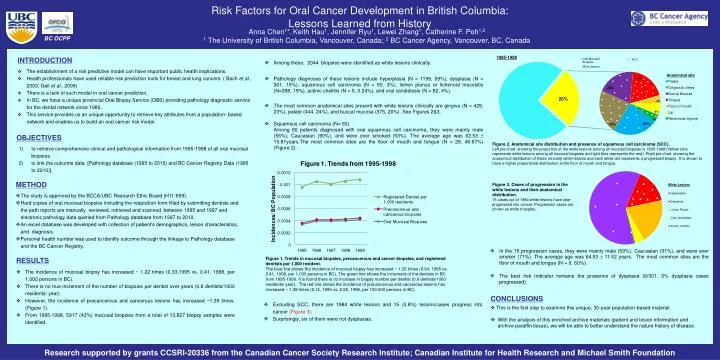
Risk Factors for Oral Cancer Development in British Columbia: Lessons Learned from History Anna Chen1*,Keith Hau1, Jennifer Ryu1, Lewei Zhang1, Catherine F. Poh1,2 1 The University of British Columbia, Vancouver, Canada; 2 BC Cancer Agency, Vancouver, BC, Canada BC OCPP • INTRODUCTION • The establishment of a risk predictive model can have important public health implications. • Health professionals have used reliable risk prediction tools for breast and lung cancers. ( Bach et al., 2003; Gail et al., 2009) • There is a lack of such model in oral cancer prediction. • In BC, we have a unique provincial Oral Biopsy Service (OBS) providing pathology diagnostic service for the dental network since 1980. • This service provides us an unique opportunity to retrieve key attributes from a population- based network and enables us to build an oral cancer risk model. • Among these, 2044 biopsies were identified as white lesions clinically. • Pathology diagnoses of these lesions include hyperplasia (N = 1198, 59%), dysplasia (N = 301, 15%), squamous cell carcinoma (N = 60, 3%), lichen planus or lichenoid mucositis (N=398, 19%), actinic cheilitis (N = 5, 0.24%), and oral candidiasis (N = 82, 4%). • The most common anatomical sites present with white lesions clinically are gingiva (N = 429, 23%), palate (444, 24%), and buccal mucosa (375, 20%). See Figures 2&3. • Squamous cell carcinoma (N= 60) • Among 60 patients diagnosed with oral squamous cell carcinoma, they were mainly male (56%), Caucasian (90%), and were ever smoked (53%). The average age was 62.53 ± 15.81years.The most common sites are the floor of mouth and tongue (N = 28, 46.67%)(Figure 2). Anatomical site 26% 4% Figure 3. Cases of progression in the white lesions and their anatomical distribution. 15 cases out of 1984 white lesions have later progressed into cancer. Progression cases are shown as white triangles. 6% 4% OBJECTIVES • to retrieve comprehensive clinical and pathological information from 1995-1998 of all oral mucosal biopsies. • 2) to link the outcome data [Pathology database (1995 to 2010) and BC Cancer Registry Data (1995 to 2010)]. Figure 2. Anatomical site distribution and presence of squamous cell carcinoma (SCC). Left pie chart: showing the proportion of the white lesions among all mucosal biopsies in 1995-1998 (Yellow slice represents white lesions among all mucosal biopsies and light blue represents the rest). Right pie chart: showing the anatomical distribution of these clinically white lesions and each white dot represents a progressed biopsy. It is shown to have a higher proportional distribution at the floor of mouth and tongue. • METHOD • The study is approved by the BCCA/UBC Research Ethic Board (H11-659). • Hard copies of oral mucosal biopsies including the requisition form filled by submitting dentists and the path reports are manually reviewed, retrieved and scanned between 1995 and 1997 and electronic pathology data queried from Pathology database from 1997 to 2010. • An excel database was developed with collection of patient’s demographics, lesion characteristics, and diagnosis. • Personal health number was used to identify outcome through the linkage to Pathology database and the BC Cancer Registry. • In the 15 progression cases, they were mainly male (53%), Caucasian (91%), and were ever smoker (71%). The average age was 64.53 ± 11.52 years. The most common sites are the floor of mouth and tongue (N = 9, 60%). • The best risk indicator remains the presence of dysplasia (9/301, 3% dysplasia cases progressed). Figure 1. Trends in mucosal biopsies, precancerous and cancer biopsies, and registered dentists per 1,000 resident. The blue line shows the incidence of mucosal biopsy has increased ~ 1.23 times (0.34, 1995 vs. 0.41, 1998, per 1,000 persons in BC). The green line shows the increment of the dentists in BC from 1995-1999. It is found there is no increase in biopsy number per dentist (0.6 dentists/1000 residents/ year). The red line shows the incidence of precancerous and cancerous lesions has increased ~ 1.39 times (2.14, 1995 vs. 2.92, 1998, per 100,000 persons in BC). • RESULTS • The incidence of mucosal biopsy has increased ~ 1.22 times (0.33,1995 vs. 0.41, 1998, per 1,000 persons in BC). • There is no true increment of the number of biopsies per dentist over years (0.6 dentists/1000 residents/ year). • However, the incidence of precancerous and cancerous lesions has increased ~1.39 times. (Figure 1). • From 1995-1998, 5917 (43%) mucosal biopsies from a total of 13,827 biopsy samples were identified. CONCLUSIONS • This is the first step to examine this unique, 30-year population-based material. • With the analysis of this enriched archive materials (patient and lesion information and archive paraffin tissue), we will be able to better understand the nature history of disease. • Excluding SCC, there are 1984 white lesions and 15 (0.8%) lesions/cases progress into cancer (Figure 3). • Surprisingly, six of them were not dysplasias. Research supported by grants CCSRI-20336 from the Canadian Cancer Society Research Institute; Canadian Institute for Health Research and Michael Smith Foundation