Download
1 / 16
170 likes | 296 Views
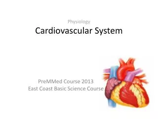
Chapter 12: The Cardiovascular System—The Heart. Function of the heart. Cells depend on interstitial fluid for survival The circulatory system balances the contents of the interstitial fluid (gas exchange, nutrients, waste, etc…)

E N D
Function of the heart • Cells depend on interstitial fluid for survival • The circulatory system balances the contents of the interstitial fluid (gas exchange, nutrients, waste, etc…) • The heart provides the mechanical “pumping” necessary for blood to circulate • Two circuits: • Pulmonary (b/t heart & lungs) • Systemic (b/t heart & body)
Anatomy of the heart • Size: 5” x 3.5” (~fist) • Shape: blunt cone • Apex: pointed end • Base: uppermost part • Location: center of thorax, behind the sternum (mediastinum) • 4 Chambers • Right atrium • Right ventricle • Left atrium • Left ventricle
Pericardial cavity (Fig. 12-2) • Pericardium encloses the heart (fist in balloon) and has 2 layers • Visceral (epicardium): inner layer closest to the surface of the heart • Parietal: outer layer • Pericardial fluid fills the cavity (lubricant)
Surface Anatomy of the heart (Fig. 12-3) • Auricle: outer flap of deflated atrium • Coronary Sulcus: groove between atria and ventricles • Anterior/Posterior Interventricular Sulci: boundary b/t lft. and rt. ventricles
The heart wall (Fig. 12-4) • Three layers • Epicardium: outermost layer (serous) • Myocardium: cardiac muscular layer; concentric wrapping • Endocardium: innermost layer, continuous with vessel linings
Internal anatomy of the heart • Interatrial Septum: separates left/right atria • Interventricular septum: separates left/right ventricles • Left/Right Atrioventricular (AV) valve • Bicuspid: left AV has 2 cusps, aka mitral valve • Tricuspid right AV has 3 cusps • Superior Vena Cava: blood from head, neck, upper limbs, and chest • Inferior Vena Cava: blood from rest of the trunk, viscera, and lower limbs • Coronary Sinus: opens into right atrium, receives blood from coronary veins
Internal anatomy of the heart • Chordae Tendineae: connects cusps to papillary muscles • Pulmonary semilunar valve: b/t right ventricle and pulmonary arteries • Aortic semilunar valve: b/t left ventricle and aorta • Aorta: start of systemic circuit • Pulmonary trunk: start of pulmonary circuit • Left/Right Pulmonary veins: receives deoxygenated blood from lungs
Heart valves (Fig. 12-6) • Pulmonary/Aortic Semilunar • No need for supportive fibers since arterial walls do not contract • Cusps of valve act like a tripod • Left/Right AV • Ventricles relaxed chordae tendineae loose & papillary muscles relaxed • Ventricles contracted chordae tendineae tense & papillary muscles contracted
Clinical note: mitral valve prolapse • Abnormalities in valve shape can prevent the valves from closing completely • Regurgitation (backflow) of blood occurs creating a soft sound (heart murmur) • Treatments: most not necessary; surgery and medication
Cardiac blood supply • Coronary circulation: blood supply to the heart muscles • Coronary arteries • Origin at the aorta (aortic sinuses) • Left (LCA) & Right (RCA) • Coronary veins • Great, Middle & Small veins • Drain into the coronary sinus (right atrium) • Myocardial infarctions (heart attacks) result from blocks in these vessels
The heartbeat Contractile Cells Conducting System Automaticity of heart contractions controlled by non-contracting cardiac muscle cells that initiate and distribute electrical impulses Sinoatrial (SA) & Atrioventricular(AV) nodes contain nodal cells (pacemaker cells @ SA node) AV bundle, bundle branches & Purkinje fibers contain conducting cells • 99% of cardiac muscle cells • Action potential in cardiac muscle • Rapid depolarization: Na+ in • The plateau: extracellular Ca2+ in • Repolarization: K+ and Ca2+out • Longer contraction and refractory than skeletal muscle
Clinical note: abnormal heart rate • Bradycardia: slow heart rate (<60bpm) • Tachycardia: fast heart rate (>100 bpm) • Arrhythmias: abnormal patterns of cardiac activity • Severe cases can be treated with defibrillator
The cardiac cycle • Heart Sounds: • Lubb: AV valves close & semilunar valves open • Dupp: ventricles relax and semilunar valves close • ECG (EKG): recording of electrical impulses in the heart • P wave: atria depolarize • QRS complex: ventricles depolarize • T wave: ventricles repolarize • The period between the start of one heartbeat and the start of the next • Systole: chamber contracts • Diastole: chamber relaxes, fills with blood and prepares for next contraction • Since blood flows from high to low pressure, ventricles are ~70% filled before atrial systole
Ecg analysis Each one of the figures represents an ECG pattern displaying three types of abnormal rhythms: Tachycardia, Bradycardia, and Arrhythmia. Identify each.