Download
1 / 57
620 likes | 915 Views
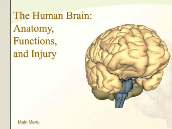
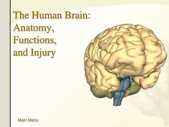
The Human Brain: Anatomy, Functions, and Injury. Main Menu. Brain Anatomy Brain Functions Injury Mechanisms. Brain Anatomy Menu. The Limbic System Cerebellum Thalamus Hypothalamus The Medulla Oblongata The Pons The Ventricles Cerebrospinal Fluid The Brainstem Brainstem Components

E N D
Main Menu • Brain Anatomy • Brain Functions • Injury Mechanisms
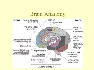
Brain Anatomy Menu The Limbic System Cerebellum Thalamus Hypothalamus The Medulla Oblongata The Pons The Ventricles Cerebrospinal Fluid The Brainstem Brainstem Components Brainstem Divisions The Cranial Nerves Skull Anatomy Interior Skull Surface Blood Vessels of the Brain Arteries of the Brain The Neuron The Meninges External Brain Structures The Cerebrum The Cerebrum – The Cortex The Neocortex Lobes of the Cerebrum Frontal Lobe Temporal Lobe Parietal Lobe Occipital Lobe Limbic Lobe
Skull Anatomy The skull is a rounded layer of bone designed to protect the brain from penetrating injuries. Blood Vessels of the Skull Rough Interior of Skull
Interior Skull Surface The base of the skull is rough, with many bony protuberances. These ridges can result in injury to the temporal lobe of the brain during rapid acceleration. Bony ridges Injury from contact with skull
Blood Vessels of the Skull The brain requires a rich blood supply, and the space between the skull and cerebrum contains many blood vessels. These blood vessels can be ruptured during trauma, resulting in bleeding. Groove for middle meningeal artery
Arteries of the Brain The human brain requires a constant supply of oxygen. A lack of oxygen of just a few minutes results in irreversible damage to the brain.
The Neuron Dendrites: Collects information from other neurons. Cell Body Axon: Transmits information to other neurons. Click image to play or pause video
The Meninges The meninges are layers of tissue that separate the skull and the brain. Skull Dura mater Arachnoid Layer Pia Mater Brain
NERVOUS SYSTEM CNS (central nervous system) Brain Spinal Cord PNS (peripheral nervous system) Peripheral Nerve Ganglion
The Cerebrum The largest portion of the brain is the cerebrum. It consists of two hemispheres that are connected together at the corpus callosum. The cerebrum is often divided into five lobes that are responsible for different brain functions. Corpus callosum
The Cerebrum Neocortex The cerebrum’s surface—the neocortex—is convoluted into hundreds of folds. The neocortex is where all the higher brain functions take place.
The Neocortex The cerebral cortex is a thin layer of cells about 1.5 to 4 mm thick. The cortex provides the connections and pathways for the highest cognitive functions, such as language and abstract thinking. The cerebral cortex contains about 25 billion neurons, more than 62,000 miles of axons, and 300,000,000,000,000 synapses. Neocortex layer The thin layer of the neocortex is dense with neurons.
Lobes of the Cerebrum Limbic Lobe Frontal Lobe Parietal Lobe Occipital Lobe Temporal Lobe
Frontal Lobe • The frontal lobe is the area of the brain responsible for higher cognitive functions. • These include: • Problem solving • Spontaneity • Memory • Language • Motivation • Judgment • Impulse control • Social and sexual behavior.
Temporal Lobe The temporal lobe plays a role in emotions, and is also responsible for smelling, tasting, perception, memory, understanding music, aggressiveness, and sexual behavior. The temporal lobe also contains the language area of the brain.
Parietal Lobe The parietal lobe plays a role in our sensations of touch, smell, and taste. It also processes sensory and spatial awareness, and is a key component in eye-hand co-ordination and arm movement. The parietal lobe also contains a specialized area called Wernicke’s area that is responsible for matching written words with the sound of spoken speech.
Occipital Lobe The occipital lobe is at the rear of the brain and controls vision and recognition.
Limbic Lobe The limbic lobe is located deep in the brain, and makes up the limbic system.
The Limbic System The limbic system is the area of the brain that regulates emotion and memory. It directly connects the lower and higher brain functions. • Cingulate gyrus • Fornix • Anterior thalamic nuclei • Hypothalamus • Amygdaloid nucleus • Hippocampus
Cerebellum The cerebellum is connected to the brainstem, and is the center for body movement and balance. Click image to play or pause video
Thalamus Thalamus means “inner room” in Greek, as it sits deep in the brain at the top of the brainstem. The thalamus is called the gateway to the cerebral cortex, as nearly all sensory inputs pass through it to the higher levels of the brain.
Hypothalamus • The hypothalamus sits under the thalamus at the top of the brainstem. Although the hypothalamus is small, it controls many critical bodily functions: • Controls autonomic nervous system • Center for emotional response and behavior • Regulates body temperature • Regulates food intake • Regulates water balance and thirst • Controls sleep-wake cycles • Controls endocrine system The hypothalamus is shaded blue. The pituitary gland extends from the hypothalamus.
The Medulla Oblongata The medulla oblongata merges seamlessly with the spinal cord and creates the base of the brainstem. The medulla is primarily a control center for vital involuntary reflexes such as swallowing, vomiting, sneezing, coughing, and regulation of cardiovascular and respiratory activity. The medulla is also the origin of many cranial nerves.
The Pons The pons is the rounded brainstem region between the midbrain and the medulla oblongata. In fact, pons means “bridge” in Latin. The main function of the pons is to connect the cerebellum to the rest of the brain and to modify the respiratory output of the medulla. The pons is the origin of several cranial nerves.
The Ventricles The ventricles are a complex series of spaces and tunnels through the center of the brain. The ventricles secrete cerebrospinal fluid, which suspends the brain in the skull. The ventricles also provide a route for chemical messengers that are widely distributed through the central nervous system. Click image to play or pause video
Cerebrospinal Fluid Cerebrospinal fluid is a colorless liquid that bathes the brain and spine. It is formed within the ventricles of the brain, and it circulates throughout the central nervous system. Cerebrospinal fluid fills the ventricles and meninges, allowing the brain to “float” within the skull. Click image to play or pause video
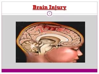
The Brainstem The brainstem is the most primitive part of the brain and controls the basic functions of life: breathing, heart rate, swallowing, reflexes to sight or sound, sweating, blood pressure, sleep, and balance. The brainstem can be divided into three major sections. Detailed brainstem anatomy. Click image to play or pause video
Brainstem Components Front More Information: Medulla Thalamus Pons Rear
Brainstem Divisions Midbrain Pons Medulla Oblongata
The Cranial Nerves • Olfactory nerve • Optic nerve • Oculomotor nerve • Trochlear nerve • Trigeminal nerve • Abducens nerve • Facial nerve • Vestibulocochlear nerve • Glossopharyngeal nerve • Vagus nerve • Accessory nerve • Hypoglossal nerve
Injury Mechanisms • The brain is a complex and delicate organ, and one that is vulnerable to injury from a variety of different traumas. These include: • Frontal Lobe Injury • Occipital Lobe Injury • Temporal Lobe Injury • Side Impact Injury • Coup/Contre-coup Injury • Diffuse Axonal Injury • Epidural Hematoma • Subdural Hematoma
Frontal Lobe Injury The frontal lobe of the brain can be injured from direct impact on the front of the head. During impact, the brain tissue is accelerated forward into the bony skull. This can cause bruising of the brain tissue and tearing of blood vessels. Frontal lobe injuries can cause changes in personality, as well as many different kinds of disturbances in cognition and memory. Click image to play or pause video
Occipital Lobe Injury Occipital lobe injuries occur from blows to the back of the head. This can cause bruising of the brain tissue and tearing of blood vessels. These injuries can result in vision problems or even blindness. Click image to play or pause video
Temporal Lobe Injury The temporal lobe of the brain is vulnerable to injury from impacts of the front of the head. The temporal lobe lies upon the bony ridges of the inside of the skull, and rapid acceleration can cause the brain tissue to smash into the bone, causing tissue damage or bleeding. Click image to play or pause video
Side Impact Injury Injuries to the right or left side of the brain can occur from injuries to the side of the head. Injuries to this part of the brain can result in language or speech difficulties, and sensory or motor problems. Click image to play or pause video
Coup/Contre-coup Injury A French phrase that describes bruises that occur at two sites in the brain. When the head is struck, the impact causes the brain to bump the opposite side of the skull. Damage occurs at the area of impact and on the opposite side of the brain. Click image to play or pause video
Diffuse Axonal Injury Brain injury does not require a direct head impact. During rapid acceleration of the head, some parts of the brain can move separately from other parts. This type of motion creates shear forces that can destroy axons necessary for brain functioning. These shear forces can stretch the nerve bundles of the brain. More on diffuse axonal injury. Click image to play or pause video
Diffuse Axonal Injury The brain is a complex network of interconnections. Critical nerve tracts can be sheared and stressed during an acceleration-type of injury. Diffuse axonal injury is a very serious injury, as it directly impacts the major pathways of the brain.
Epidural Hematoma An epidural hematoma is a blood clot that forms between the skull and the top lining of the brain (dura). This blood clot can cause fast changes in the pressure inside the brain. When the brain tissue is compressed, it can quickly result in compromised blood flow and neuron damage. Click image to play or pause video
Subdural Hematoma A subdural hematoma is a blood clot that forms between the dura and the brain tissue. The clot may cause increased pressure and may need to be removed surgically. When the brain tissue is compressed, it can quickly result in compromised blood flow and tissue damage. Click image to play or pause video
Brain Functions • Vision • Taste • Cognition • Emotion • Speech • Language • Hearing • Motor Cortex • Sensory Cortex • Autonomic Functions
Vision The visual cortex resides in the occipital lobe of the brain. Sensory impulses travel from the eyes via the optic nerve to the visual cortex. Damage to the visual cortex can result in blindness.
Taste The gustatory complex (green circle) is the part of the sensory cortex (purple area) that is responsible for taste.
Cognition The prefrontal cortex is involved with intellect, complex learning, and personality. Injuries to the front lobe can cause mental and personality changes.
Emotion Prefrontal cortex Emotions are an extremely complex brain function. The emotional core of the brain is the limbic system. This is where senses and awareness are first processed in the brain. Mood and personality are mediated through the prefrontal cortex. This part of the brain is the center of higher cognitive and emotional functions. Limbic system