Download
1 / 1
10 likes | 176 Views
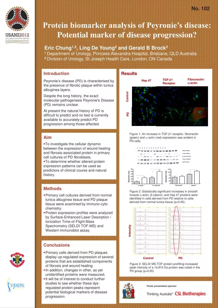
C C P P P C P C C P P P P. Tubulin (42 kDa). a - actin (40 kDA). b - catenin (92 kDa). TGTase II (85 kDa). Hsp47 (47 kDa). No. 102. Protein biomarker analysis of Peyronie’s disease: Potential marker of disease progression?.

E N D
C C P PP C P C C P PPP Tubulin (42 kDa) a-actin (40 kDA) b-catenin (92 kDa) TGTase II (85 kDa) Hsp47 (47 kDa) No. 102 Protein biomarker analysis of Peyronie’s disease: Potential marker of disease progression? Eric Chung1,2, Ling De Young2 and Gerald B Brock2 1 Department of Urology, Princess Alexandra Hospital, Brisbane, QLD Australia 2 Division of Urology, St Joseph Health Care, London, ON Canada Introduction Peyronie’s disease (PD) is characterised by the presence of fibrotic plaque within tunica albuginea layers. Despite the long history, the exact molecular pathogenesis Peyronie's Disease (PD) remains unclear. At present the natural history of PD is difficult to predict and no test is currently available to accurately predict PD progression among those affected. Results Fibronectin/ -actin TGF-1 Receptor Hsp 47 Control PD Figure 1: An increase in TGF-1 receptor, fibronectin (green) and -actin (red) expression was evident in PD cells. • Aim • To investigate the cellular dynamic between the expression of wound healing and fibrosis-associated protein in primary cell cultures of PD fibroblasts. • To determine whether altered protein expression patterns can be used as predictors of clinical course and natural history. • Methods • Primary cell cultures derived from normal tunica albuginea tissue and PD plaque tissue were examined by immuno-cyto-chemistry. • Protein expression profiles were analyzed by Surface-Enhanced Laser Desorption / Ionization Time-of-Flight Mass Spectrometry (SELDI TOF-MS) and Western immunoblot assay. Figure 2: Statistically significant increases in smooth muscle -actin, β-catenin, and Hsp 47 proteins were identified in cells derived from PD relative to cells derived from normal tunica tissue (p<0.05). Intensity • Conclusions • Primary cells derived from PD plaques display up-regulated expression of several proteins that are established components of fibrosis and wound healing. • In addition, changes in other, as yet unidentified proteins were measured. • It will be of interest to conduct further studies to see whether these dys-regulated protein peaks represent potential biological markers of disease progression. Control PD Figure 3: SELDI MS-TOF protein profilling increased peak intensity of a 10,810 Da protein was noted in the PD group (p<0.05) Poster presentation sponsor