Download
1 / 47
470 likes | 683 Views
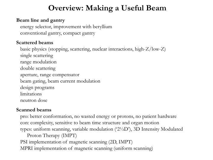
Overview: Making a Useful Beam. Beam line and gantry energy selector, improvement with beryllium conventional gantry, compact gantry Scattered beams basic physics (stopping, scattering, nuclear interactions, high-Z/low-Z) single scattering range modulation

E N D
Overview: Making a Useful Beam Beam line and gantry energy selector, improvement with beryllium conventional gantry, compact gantry Scattered beams basic physics (stopping, scattering, nuclear interactions, high-Z/low-Z) single scattering range modulation double scattering aperture, range compensator beam gating, beam current modulation design programs limitations neutron dose Scanned beams pro: better conformation, no wasted energy or protons, no patient hardware con: complexity, sensitive to beam time structure and organ motion types: uniform scanning, variable modulation (‘2½D’), 3D Intensity Modulated Proton Therapy (IMPT) PSI implementation of magnetic scanning (2D, IMPT) MPRI implementation of magnetic scanning (uniform scanning)
Beam Line at the Burr Center Cyclotron-based therapy facilities use degraders to change the beam energy. (Variable-energy cyclotrons are very expensive and energy changes are very slow.) The resulting increase in energy and angular spread is trimmed away by slits, but a lot of beam is lost (up to 99%). The energy selector system is not needed in a synchrotron based facility.
Choice of Energy Degrader Material An energy selector system (ESS) has very low beam transmission (≈1% or less) when set up for low energies, because the spread in energy and angle introduced by the thick degrader cannot be transported by the magnetic beam line. That can be improved somewhat by using a lower-Z degrader material. These data (Gottschalk and Wagner, HCL technical note 11/16/99) show a 40% increase obtained by using a beryllium degrader instead of the stepped carbon wedge. Beryllium is shunned at some labs because of potential hazards. It is used in the ESS at the Indiana Univ. Cyclotron Facility (IUCF).
Conventional Gantry Framework of the IBA gantry. This is a massive structure, three stories high. It carries the final section of the beam line and the beam spreading ‘nozzle’ so that beam can be directed at the patient from any direction. It strives for sub-mm accuracy in the center of rotation, a major challenge to the mechanical engineer. What the patient sees: gantry room at the Midwest Proton Radiotherapy Institute (MPRI). This is a modified IBA gantry. Also visible are an industrial robot adapted to serve as the patient couch, the proton ‘snout’ which carries the aperture and range compensator, and an X-ray imaging plate used during patient alignment.
Compact Gantry (Paul Scherrer Institute, PSI) This gantry at PSI near Zurich, Switzerland, has a novel design to fit into the available space. The patient couch counter-rotates with the large bending magnet, so when treated from below the patient is up in the air. One scanning dimension is provided by a sweeping magnet just before the 90º bend, the second (depth) by degraders just upstream of the patient and the third by couch motion. The system, operating since 1996, is used for variable modulation or for IMPT. It has recently been upgraded with a dedicated superconducting cyclotron (ACCEL Corp., a division of Varian) and a full-sized gantry with spot scanning is planned (‘PROSCAN’ project).
Stopping In matter, protons slow down and stop by millions of collisions with atomic electrons. If we measure range by mass traversed rather than distance, the range in air is similar to water. If we place a stack of material just upstream of a charge collector we find that protons of a definite energy have a characteristic range, defined as the halfway point of the steep falloff. However, even that has some width: the protons do not all stop at exactly the same depth. This range straggling spread is roughly 1.2% of the range itself and depends very little on the stopping material. The slow falloff is caused by loss of primary protons to nuclear reactions. The upper curve obtains if we move the Faraday Cup closer, allowing it to catch more of the wide-angle charged nuclear secondaries.
Stopping Theory A very successful theory of stopping was developed by Bethe and others in the 1930’s. However, the stopping power of each material depends on an empirical parameter, the mean ionization energy I for that material. Because I is difficult to choose, we usually rely on standard tables such as ICRU Report 49 instead of computing stopping power directly from the theory. It turns out that range is nearly proportional to energy on a log-log plot (above) which implies an approximate power law R ≈ aTb . The energy range of clinical relevance is 3-300 MeV .
Multiple Scattering L x0 θ0 MP When protons pass through a slab of material they suffer millions of collisions with atomic nuclei. The statistical resultant is a multiple scattering angle whose distribution is approximately Gaussian. For protons, this angle is always small so the projected displacement in any measuring plane (MP) is also Gaussian. The width parameter of the angular distribution is θ0 . The corresponding displacement x0 can easily be measured by scanning a dosimeter across the MP. The task of multiple scattering theory is to predict θ0 given the scattering material, thickness and incident proton energy.
Multiple Scattering Theory The definitive theory of multiple Coulomb scattering was published by G. Molière in 1947. It has no empirical parameters and (very important for proton therapy design) covers arbitrarily thick scatterers (that is, the effect of energy loss) as well as compounds and mixtures. The angular distribution at large angles falls off roughly as 1/θ4 but is nearly Gaussian for small angles, a good enough approximation for most proton therapy calculations. θ0 , the width parameter of the Gaussian, is given by Molière theory as shown by this figure (Gottschalk et al. NIM 74 (1993) 467). The points are experimental. Molière theory is fairly difficult to evaluate but an excellent and simple approximation to it, Highland’s formula, exists.
Nuclear Reactions About 20% of 160 MeV protons stopping in water have a non-elastic nuclear reaction where the primary proton is seriously degraded and secondary protons, neutrons and nuclear fragments appear. This figure (M. Berger, NISTIR 5226 (1993)) shows the effect on the Bragg peak. Dose from the EM peak shifts upstream, lowering the peak and flattening the entrance region especially at high proton energy. Because nuclear reactions are rather hard to model, our programs take them into account by simply using measured rather than predicted Bragg peaks for the depth-dose dependence.
High-Z/Low-Z Energy loss and multiple scattering for 160 MeV protons incident on 1 g/cm2 of various materials; LR ≡ radiation length.. For scattering with minimum energy loss, use lead. To reduce energy with minimum scattering, use beryllium.
Single Scattering The simplest treatment beam. With enough scatterer, we can make the Gaussian at the target wide enough so it is sufficiently flat (say 5% or ±2.5%) over the required radius. Unfortunately, only 5% of the protons will fall within that radius. Also, the scatterer uses up a lot of the proton energy. To treat large or deep fields we need something better. Single scattering is used for targets, like the eye, which are small and shallow.
Range Shifter and Modulator At a fixed energy machine we may need some extra material to bring the dose forward to the desired distal depth. Also, if the target has significant extent in depth we can use a modulator wheel of different thicknesses of plastic to sweep the proton endpoint back and forth. The dwell time of each step in the beam is carefully computed so the resulting spread out Bragg peak (SOBP) will be flat.
Double Scattering For better efficiency (up to 45%) and less energy loss we use double scattering. A flat first scatterer produces a Gaussian on the second scatterer, which is stronger in the center to scatter those protons more. With careful design the transverse dose at the target is flat. A complementary plastic plate is used to compensate energy loss across S2. The first scatterer can be a simple sheet, or it can be a modulator wheel as shown.
Patient-Specific Hardware Having made a large uniform cylindrical dose field, we shape it as required for that particular target. The patient apertureAP blocks protons outside the target cross section. A range compensatorRC shapes the distal dose surface and can also compensate for inhomogeneities is the patient. The snoutSN holds this hardware while blocking unwanted large-angle protons. A therapy center should have snouts of several sizes.
Beam Gating, Beam Current Modulation Without a few tricks we would need a very large library of modulators to cover the range of clinical requirements, because a given mod only works well over a small range of incident energy. The first trick is beam gating. A wheel or track designed for full mod can be used for anything less by just turning off the beam during the unwanted proximal steps. Disadvantages: a slight rounding of the proximal corner (shown above) because the beam usually covers several steps, some decrease in dose rate, and additional equipment and QA because we need to know the exact wheel angle at every instant. The second trick is beam current modulation: adjusting the beam current as a function of time to correct small errors in the SOBP. Both techniques are used in the standard IBA nozzle. Just three or four basic modulator tracks can cover the whole range of requirements in one room.
Design Programs Although we can design scattering systems accurately from first principles the math is complicated and we need special programs like NEU(by the author). This run shows an experimental test of the first ‘upstream modulator’ double scattering system ca. 1990 at the Harvard Cyclotron Lab. Except for the transverse width the data agree rather well.
Limitations Passive beam spreading is limited by the fact that nature does not provide perfect scatterers (no energy loss) or degraders (no scattering). The limits are best visualized by graphing the attainable field radius vs. maximum treatment depth. In this case we assume 230 MeV protons, a throw of 250 cm and S2 placed at 50 cm. The relative modulation (mod/depth) obtainable at each point is indicated by the degree of filling of the point. Solid means full modulation or anything less. A larger throw would yield deeper fields for the same energy.
Unwanted Neutron Dose In addition to unwanted proton dose patients receive some neutron dose: unavoidable internal neutrons from therapy protons interacting in the target plus external neutrons from the beam spreading system. These originate wherever a lot of protons lose a lot of energy (highlighted above). In a well designed scattering system the main source is the patient collimator, so we should match the open field size to the target, and the external neutron dose is comparable to the internal dose. Many studies show that both are on the order of 1mSv/Gy. The attributable lifetime risk of a fatal cancer is <1% for a mixed population.
Magnetic Scanning: Advantages The following applies specifically to the PSI ‘point and shoot system’ (Pedroni et al., Med. Phys. 22 (1995) 37) but most points are valid for scanning generally. Better conformation to the target: spot scanning gives the best ratio of target dose to unwanted dose, eliminating for example the overdosing from fixed modulation. The reduction in integral proton dose is ≈10% (Goitein and Chen, Med. Phys. 10 (1983) 831, Urie and Goitein, Med. Phys. 16 (1989) 593) Patient-specific hardware is optional: could be a considerable operational advantage. However, spot scanning is backward compatible with apertures or range compensators should these still be desirable, e.g. shallow targets. More efficient beam and energy utilization: all protons are used, reducing unwanted neutron dose and accelerator activation. No beam energy is wasted in scatterers. Compact gantry: the throw of the PSI spot scanning system is much shorter than a conventional gantry. (Note, however, that PSI plans a full sized gantry with a conventional patient couch for their current upgrade ‘PROSCAN’.) Simplifies field patching: no inherent limitation on field size. Beam gating easy to implement: all the hardware is already there.
Magnetic Scanning: Drawbacks Sensitive to accelerator time structure: the DC beam from a cyclotron is ideal for spot scanning. Coordinating scanning with the pulsed output of a synchrotron raises additional problems, which have, however, been tackled successfully at GSI. Organ motion: because of practical limits on magnetic sweep speeds, a moderately large target can only be painted once per treatment session. Therefore target motion can lead to serious under- or over-dosing. More complex technical infrastructure required: Spot scanning has so far been realized only at a few centers: PSI (Zurich), GSI (Darmstadt), M.D. Anderson (Houston), Rinecker Center (Munich), HIT (Heidelberg). It requires greater technical and financial resources.
Magnetic Scanning: Strategies Uniform scanning: once called ‘wobbling’. A system using a rotating 1.6 kG permanent magnet followed by a Pb scatterer was tested at Harvard (Koehler et al., Med. Phys. 4 (1977) 297) but never used in clinical practice. The same article mentions a ‘pair of sweeping magnets’ used at Uppsala. The object is to get a reasonably large field with good efficiency without the energy loss from scatterers. A modern version at MPRI with a combined X-Y scanning magnet is described below. Efficiency is only slightly better than passive spreading, and patient-specific hardware is still needed. Variable modulation: sometimes called ‘2½D’. A relatively small number of uniform fields are combined as one would in passive spreading, but with variable range modulation during the scan to reduce integral dose and dose to organs at risk. Patient hardware is optional. Most of the treatments at PSI are of this nature. IMPT: a number of non-uniform fields are combined to produce a uniform dose (if desired) in the target. This affords the maximum flexibility, the best dose conformation, and also the greatest complexity in planning and treatment delivery. Local or remote energy variation: early systems used degraders between the magnet and the patient to achieve range modulation (Bragg peak stacking). That is simple but has the disadvantages of degraders: scattering and neutron production. A ‘pure’ scanning system varies the beam energy out of the accelerator, and the entire beam line must be retuned rapidly (Rinecker, MD Anderson, Heidelberg).
Spot Scanning at PSI (Zurich) This facility has operated since 1996 treating some 30 patients/year. A major upgrade including a dedicated superconducting cyclotron is in progress and recent patient numbers should be up. In the original version a low-current parasitic beam, up to 214 MeV, is obtained from the 590 MeV cyclotron. The scenario is ‘point and shoot’. An upstream beam kicker magnet lets the beam be turned on and off quickly. A 1D sweeper magnet just upstream of the 90º bend in the compact gantry provides the fastest axis of scanning. The second fastest is depth, provided by degrader plates just upstream of the patient. The third and slowest axis is couch motion. The system is described by Pedroni et al., Med. Phys. 22 (1995) 37 (their Figure 3 is at left). Treating a 1 liter volume requires about 10,000 spots delivered in 3.5 minutes, about half of that being deadtime.
PSI: Spot Scanning with a Compact Gantry Moving with the beam: sweeper magnet (red); 90º 1D bend magnet (blue); strip ion chambers to measure position and total dose per spot (yellow); 36 polyethylene plates, each 4.7mm water equivalent, plus one half-thickness plate (green). Each plate can be moved in pneumatically (30ms) but they must all be moved out together (200ms). The couch counter-rotates with the magnet. Couch motion is the slowest axis.
Cheating Nature With spot scanning one can actually make the dose falloff sharper (compared with a collimator) for deep targets where multiple scattering in water dominates. By spacing spots carefully one can get a dropoff which is closer to that of the underlying Gaussian than to the error function, increasing dose gradient by about 1.6× with a negligible sacrifice of dose uniformity. The method is analogous to what one does at the distal edge of a range-modulated depth-dose distribution. The spot application system uses this strategy automatically. (Figure from Pedroni et al., Med. Phys. 22 (1995) 37.)
Uniform Scanning at MPRI This is the floor plan of the Midwest Proton Radiotherapy Institute (MPRI). A proton therapy center, built around the old IUCF 208 MeV cyclotron, began operations in 2003. A trunk beam line serves a fixed beam room, two gantries and a research room. Each room has its own energy selector (ESS) using beryllium wedge degraders. MPRI uses uniform scanning as a way of making fairly large fields while preserving proton energy. The gantries are modified IBA gantries. The nozzles are MPRI’s design. Industrial robots are adapted for use as patient couches.
Combined X-Y Scanning Magnet at MPRI This magnet (V. Anferov, Med. Phys. 32 (2005) 815) bends the beam in either or both directions, taking up less of the beam line than separate magnets. Also, the source distance is the same for x and y, greatly simplifying the treatment planning program.
Uniform Scanning Nozzle at MPRI The MPRI nozzle (Anferov et al, Proc. EPAC 2006, Edinburgh) combines commercial IBA components with devices designed and fabricated by IUCF. Using a computer controlled degrader array it generates an SOBP adjustable from 2 to 15cm at depths up to 27cm water. The combined X-Y magnet generates a field up to 30cm in diameter. The SOBP is measured efficiently by a multi-layer ionization chamber (not shown).
Accelerators This course does not cover accelerators but let’s do a quick overview. Conventional photon machine: a gantry mounted electron linac (≈10 MeV), 270 bend, target, flattening filters, multi-leaf collimator. Linear accelerators: do not look attractive for proton radiotherapy. Much more current than needed, fixed energy, too large and expensive with current technology. FM cyclotron: 160 MeV (HCL, Dubna) Isochronous cyclotron: 230 MeV (all IBA centers) Superconducting isochronous cyclotron: 250 MeV (Varian/ACCEL centers) Synchrotron: 250 MeV (Loma Linda, M.D. Anderson) Superconducting FM cyclotron: 250 MeV (Still River Systems) An isochronous machine cannot use the full field (≈9 Tesla) available with superconductivity because of focusing considerations. Therefore the only viable idea for a truly compact machine is a superconducting FM cyclotron, currently under development at SRS
FM Cyclotron HCL: 160 MeV, ≈1000 tons.
Isochronous Cyclotron IBA : 230 MeV, ≈200 tons.
Superconducting Isochronous Cyclotron Varian/ACCEL: 250 MeV, ≈90 tons.
Synchrotron The 250 MeV synchrotron for the Loma Linda University Medical Center under construction at Fermilab ca. 1988. The injector is an RFQ (Radio Frequency Quadrupole).
Superconducting FM Cyclotron Still River Systems: 250 MeV, 15 tons (under development)
1946 Harvard 1996 IBA 2000 Accel 2008 Still River
Next Generation Proton Therapy Still River Systems treatment room concept
As of Mar 2013, there were a total of 35 proton therapy centers in Canada, China, England, France, Germany, Italy, Japan, Korea, Poland, Russia, South Africa, Sweden, Switzerland, and USA. To date more than 83,000 patients had been treated.
Proton Therapy Centers Currently Operating in the USA Loma Linda Medical Center – James M. Slater Proton Therapy Center (1990) -Fermilab/Optivus University of California - UC Davis Proton Facility (1994) – Ocular treatments only -U. Cal. MGH – Francis H. Burr Proton Therapy Center (2001) -IBA Indiana University - Midwest Proton Radiotherapy Institute (2004) -IUCF/IBA University of Florida Proton Therapy Institute (2006) -IBA University of Texas M. D. Anderson Cancer Center (2006) -Hitachi Oklahoma ProCure Treatment Center – Oklahoma City (2009) -IBA Roberts Proton Therapy Center – University of Pennsylvania (2010) –IBA Hampton University Proton Therapy Institute - Hampton, VA (2010) –IBA Chicago ProCure Treatment Center - Warrenville, IL (2010) –IBA ProCure Proton Therapy Center – Somerset, NJ (2012) – IBA SCAA ProCure Proton Therapy Center – Seattle, WA (2013) – IBA
Proton Therapy Centers Under Construction in the USA Siteman Cancer Center - St. Louis, MO (2013) –Mevion (formerly Still River Systems) The Robert Wood Johnson University Hospital - New Brunswick, NJ (2013) - Mevion Scripps Medical Center – San Diego, CA (2013) –Varian McLaren Proton Center – Flint, MI (2013) – ProTom University of Oklahoma – Oklahoma City, OK (2013) – Mevion Provision Center for Proton Therapy – Knoxville, TN (2014) - IBA Mayo Clinic – Rochester, MN (2014) – Hitachi University of Maryland – Baltimore, MD (2014) –Varian Willis-Knighton Cancer Center – Shreveport, LA (2014) – IBA* Mayo Clinic – Phoenix, AZ (2015) – Hitachi St. Jude Hospital – Memphis, TN (2015) - Hitachi MD Anderson Cancer Center – Orlando, Fl (2015) – Mevion First Coast Oncology -Jacksonville, Fl (2015) – Mevion Emory Proton Therapy Center – Emory, GA (2016) – Varian Texas Center for Proton Therapy – Irving, TX (2016) - IBA
Summary We have discussed, very briefly, how a compact beam is delivered to the treatment room. If the proton source is a fixed energy cyclotron, an energy selection degrader and re-analysis system may be used to vary the energy into the treatment room. This is not necessary for a synchrotron, which has variable energy. The ESS has very poor transmission at energies below 100 MeV. Once in the treatment room the beam is spread out to cover the target. Passive spreading uses one or two scatterers to prepare an oversize beam which is then shaped by a patient aperture. Currently used for about 97% of proton patients, this method is simple, foolproof and relatively efficient (45%). However, it requires patient-specific hardware, wastes some proton energy, produces some excess neutrons, and does not conform the dose to the target as well as could be. Active spreading by means of magnets can overcome these problems. It has proven difficult to implement given the resources of the commercial/clinical community, but several systems are now operational (Hitachi, Varian/ACCEL). Technical and QA problems aside, the main clinical problem is organ motion, which can lead to significant dose errors because the target is usually painted only once per treatment session. A pure scanning system varies proton energy at the machine. The entire beam line must be retuned rapidly. Two older systems sidestep that problem, using degraders between the last magnet and the patient.