Download
1 / 50
500 likes | 791 Views
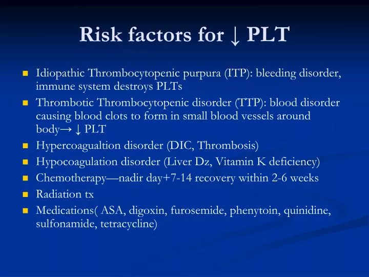
Risk factors for ↓ PLT. Idiopathic Thrombocytopenic purpura (ITP): bleeding disorder, immune system destroys PLTs Thrombotic Thrombocytopenic disorder (TTP): blood disorder causing blood clots to form in small blood vessels around body→ ↓ PLT Hypercoagualtion disorder (DIC, Thrombosis)

E N D
Risk factors for ↓ PLT • Idiopathic Thrombocytopenic purpura (ITP): bleeding disorder, immune system destroys PLTs • Thrombotic Thrombocytopenic disorder (TTP): blood disorder causing blood clots to form in small blood vessels around body→ ↓ PLT • Hypercoagualtion disorder (DIC, Thrombosis) • Hypocoagulation disorder (Liver Dz, Vitamin K deficiency) • Chemotherapy—nadir day+7-14 recovery within 2-6 weeks • Radiation tx • Medications( ASA, digoxin, furosemide, phenytoin, quinidine, sulfonamide, tetracycline)
Mgmt Thrombocytopenia • PLT transfusions PLT<10,000 • Colony stimulating factors, eg IL 11 (Neumega) • Steroids • Progesterone ↓ menstrual bleeding
Nursing interventions • Avoid tight BP cuffs when PLT<20,000 • Avoid invasive procedures • Avoid sharp objects/no barefoot • Apply firm pressure to venipuncture sites for 5 minutes • Treat nose bleeds with high fowler’s position-icepack • Prevent constipation • Encourage soft toothbrushes
Why do we evaluate WBC? • To assess body's response to significant bacterial insult, such as appendicitis, Pelvic inflammatory disease, pneumonia, pyelonephritis, and SEPSIS
WHITE BLOOD CELLS • Segmented Neutrophil (60%) • Lymphocytes(30%) • Monocytes(6%) • Eosinophils(3%) • Basophils(1%)
SEGMENTED NEUTROPHIL • 1ST line of defense • Normal 50-70% • Survive 1-2 DAYS
LYMPHOCYTES • 2 Types by appearance: Large granular and small • Large granular are NK cells • Small are T & B cell • T cell mediated immunity • B cell humoral immunity
MONOCYTE • 5-10 % Circulating WBCs • When stimulated become Macrophages and dentritic cells in the tissue • Clean up the debris
EOSINOPHIL • 1-5% Circulating WBCS • Involved in parasitic infections • Involved with mechanism associated with allergy and asthma • ↑In case of w.w.w.
BASOPHIL • < 1% circulating WBCS • Involved with allergic and inflammatory response • Release histamines and cytokines
Hematological Symptoms • Neutropenia—defined as ANC<1000/mm3 • Anemia--↓RBC & Hgb • Thrombocytopenia—low PLT <100,000 • Leukopenia—decrease in WBC, below the lower limit • Pancytopenia-an abnormal deficiency in all blood cells, RBC, WBC, & PLT; usually associated with bone marrow tumor or with aplastic anemia)
Risk factors for febrile neutropenia • Previous Hx Chemo/Type of chemo/prior radiation Tx • Age>65/ female gender • Poor nutritional status • Advanced cancer and bone marrow involvement • ↑LDH, ↓Hgb • Leukemia/lymphoma/lung cancer • Open wounds • DM • COPD
Prevention of Neutropenia • Growth stimulating factors activate production of bone marrow cells. It can be given prophylactically or therapeutically • BMT give GCSF on day+4 of stem cell SCT • GCSF-/filgrastim 5mcg/kg • Pegfilgrastim--Neulasta 6mcg/kg
Neutropenic Precautions • Limit exposure of pts to infections • Hand washing ↓spread • Avoid crowds • Avoid fresh fruits /vegetables/flowers • Avoid caring for animals esp. cleaning excretes • Avoid gardening
Infections • Mechanical barriers—skin, mucous membranes • Chemical barriers-pH of tissues • Inflammatory and immune responses
Risks of Infections • High risk • Intermediate risk • Low risk
High Risk for infection • Allogeneic BMT • Acute Leukemia's • GVHD tx ↑dose steroids • Neutropenic lasting >10 days • Break in skin/mucosal barrier • Prolonged ABX or steroid use • Poor nutrition • Invasive procedure • Poor personal hygiene
Intermediate Risks for infections • Autologous BMT • Lymphoma/MM/CLL • Neutropenic anticipated to last >7-10 days
Low Risk for infections • Standard Chemotherapy • Neutropenia <7 days
PT AND PTT • PT (PROTIME) Test the extrinsic pathway • PTT Tests the intrinsic pathway • COUMADIN (WARFARIN) Affects the Vitamin K factors (II,VII,IX,X ) of which factor VII is the most labile • Hemophilia is a factor VIII deficiency • INR: Method for standardizing Protimes. It is a ration of tested results : control
COAGULATION PRODUCTS • Fresh Frozen Plasma • Cryoprecipitate: Factor VIII, Fibrinogen • Activated Products: Factor IX
Chemistry Tests • Liver Function Studies • Renal Function • Electrolytes—Na, K, Ca, Mg, Po4, Co2
LIVER FUNCTION Hepatocelluar Enzymes: • AST (ASPARTATE AMINOTRANSFERASE) • ALT (ALANINE AMINOTRANSFERASE) • SGGT (very specific) • LDH (Lactate Dehydrogenase)
LIVER FUNCTION ALKALINE PHOSPHATASE-AP not specific to liver AP= LARGE COMPONENT IN BONE BILIRUBIN -2 TYPES DIRECT OR CONGUGATED AND INDIRECT OR UNCONGUGATED. • AP / BILIRUBIN the first enzymes to rise with liver GVHD • INDIRECT BILIRUBIN associated with hemolysis
LDH • Nearly every type of cancer, as well as many other diseases, can cause LDH levels to be ↑, cannot be used to dx a particular type of cancer. • LDH levels can be used to monitor treatment of some cancers, including testicular cancer, Ewing's sarcoma, non-Hodgkin's lymphoma, and some types of leukemia • Elevated LDH levels can be caused by a number of noncancerous conditions, including heart failure, hypothyroidism, anemia, and lung or liver disease.
RENAL • BUN (BLOOD UREA NITROGEN) Elevated with: Kidney dysfunction, Dehydration, excess protein in blood such as TPN, High protein diet, GI bleeding • Creatinine: chemical waste is generated from muscle metabolism. Creatinine is produced from creatine, a molecule of major importance for energy production in muscles. Creatinine is transported through the bloodstream to the kidneys. The kidneys filter out most of the creatinine and dispose of it in the urine.
CREATININE CONT. • The ratio of BUN : creatinine determines renal dysfunction VS pre-renal dysfunction such as dehydration. • CALCULATED CREAT. CLEARANCE: in ml/min---CRCL=(140-AGE) x ideal B.W./Scr. x 72 (x 0.85 for females)
ELECTROLYTES • SODIUM • POTASSIUM • CALCIUM • PHOSPHORUS • MAGNESIUM • CO2
SODIUM/POTASSIUM • SODIUM /POTASSIUM MEMBRANE PUMP
CALCIUM/PHOSPHORUS • DIRECT INTERACTION • IF GIVEN CONCOMBINETLY: NaPO4 + CaCO3 =
CALCIUM • Calcium in plasma is bound to Albumin. If Albumin is low, you get a falsely low serum calcium. • 2 ways to get a more accurate Calcium: IONIZED CALCIUM CORRECTED CALCIUM • CORRECTED Calcium: [(4.0 – serum alb) x 0.8 ] + s Ca = corrected Ca
MAGNESIUM • Very important for Cardiac, Nervous and GI systems. • Interacts with calcium and Potassium. • Difficult to get a Normal serum level of Potassium if Mg is low.
CO2 • Gives a rough idea of pH and buffer system in blood • Infections typically have low venous CO2
Bone marrow Biopsy-aspirate • Used in Identi. metastatic Dz, esp hematological malignancies • Assess iron stores • Assess megaloblastic maturation, in Vit B12 and folate deficiencies or in MDS • Assess fat atrophy, aplasia or fibrosis
Other tests done in Hem/Onc • Fractionated bilirubin—to differentiate cause of hyperbilirumia • Stool guaiac--? bleeding • Coombe’s test—direct/indirect • Haptoglobin level—to detect hemolytic anemia • Hgb electropheresis----
MICROBIOLOGY • BACTERIA • VIRUS • FUNGAL/YEAST • PROTOZOAN
BACTERIA • CATAGORIZED BY SHAPE AND STAINING PROPERTIES • GRAM’S STAIN • SHAPES ARE COCCI, RODS, AND SPIROCHETES
COCCI RODS SPIROCHETES BACTERIAL SHAPES
GRAM’S STAIN • INTERACTS WITH THE BACTERIAL MEMBRANE AND STAINS IT EITHER BLUE OR RED • BLUE IS GM (+) • RED IS GM (-)
GRAM POSITIVE • COCCI: 1. STAPHALOCOCCUS EITHER COAGULASE (-) OR (+) COAG NEG=STAPH EPID. COAG POS= STAPH AUREUS 2. STREP, ENTEROCOCCUS • RODS: BACILLUS, LISTERIA, CORYNEBACTERIA DIPTHERIA
GRAM NEGATIVE • COCCI: NEISSERIA, MORAXELLA, ACINETOBACTER • RODS: E.COLI, PSEUDOMONAS, SHIGELLA, SALMONELLA, KLEBSIELLA, PROTEUS, ENTEROBACTER, VIBRIO
FUNGUS • MOLDS: ASPERGILLUS , MUCOR, • YEAST AND YEAST-LIKE: CANDIDA, TORULOPSIS, HISTOPLASMOSIS, CRYPTOCOCCUS, BLASTO.
VIRUS • CYTOMEGALOVIRUS • EPSTEIN-BAR • BK • ADENOVIRUS • INFLUENZA A & B • PARAINFLUENZA • RSV
PNEUMOCYSTIS CARINII • PREVIOUSLY CONSIDERED A PROTOZOAN BUT NOW IN FUNGUS CLASS
References • Demetri, G. (2001) Anemia and its functional consequences in cancer pts, current challenges in management & prospects for improving therapy. British Journal of Cancer,84,31-37 • Dessypris, E, Erythropoiesis (1988). Lee G R, Foster J, Lukens J, Wintrobe M M, eds.Wintrobe’s clinical hematolology. Pa: Lippincott Williams & Williams 1998. 169-192. • Ludwig H, & Strasser K.(2001) Symptomatology of anemia. Seminars in oncology, 28, 7-10 • Means, R. (1999). The anemias of chronic disorders. Lee GR, Foerster J, Lukens J, Paraskeras F, Greer J P, Rodgers GM, eds. WIntrobe’s Clinical hematolgoy.10th ed, Pa. Lippincott Williams & Wilkin;1999:1011-1021 • National Comprehensive Cancer Network (2007). Cancer and treatment related anemias, version 3.2007