Download
1 / 58
580 likes | 766 Views
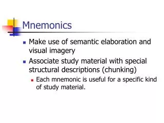
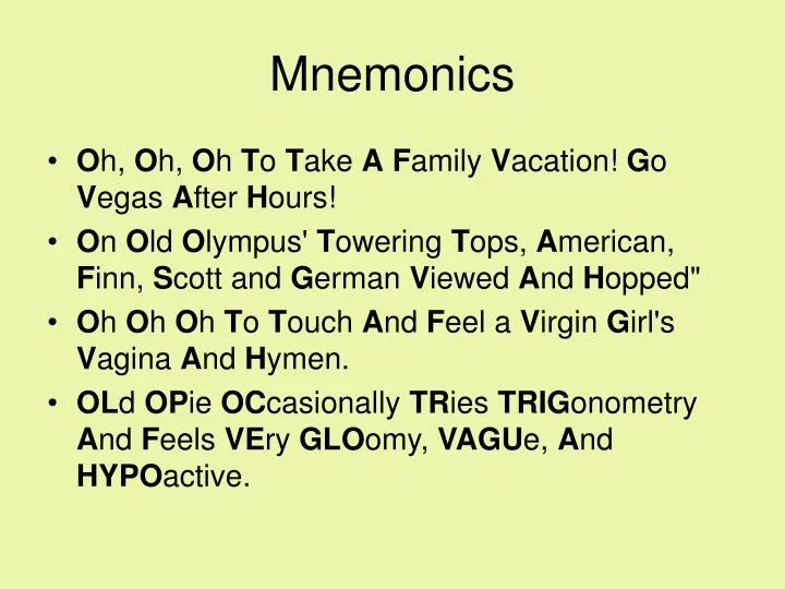
Mnemonics. O h, O h, O h T o T ake A F amily V acation! G o V egas A fter H ours! O n O ld O lympus' T owering T ops, A merican, F inn, S cott and G erman V iewed A nd H opped" O h O h O h T o T ouch A nd F eel a V irgin G irl's V agina A nd H ymen.

E N D
Mnemonics • Oh, Oh, Oh To Take AFamily Vacation! Go Vegas After Hours! • On Old Olympus' Towering Tops, American, Finn, Scott and German Viewed And Hopped" • Oh Oh Oh To Touch And Feel a Virgin Girl's Vagina And Hymen. • OLd OPie OCcasionally TRies TRIGonometry And Feels VEry GLOomy, VAGUe, And HYPOactive.
CRANIAL NERVES1st part David Kachlík
Numeral clasification • Claudius Galenus (2nd century) 7 pairs • Thomas Willis (1664) 9 pairs • Samuel Thomas von Sömmerring (1778) 12 pairs
Developmental classificationmediolaterally • somatomotor somatic • somatomotor branchial • visceromotor • viscerosensory • somatosensory • special sensory
SomatoMotor somatic CN • preotic myotoms (somitomers) form external muscles of eyeball – n. III, IV, VI • occipital somites form muscles of tongue – n. XII
Special sensory CN VIII. I. II.
Where CN emerge from brain? I. – telencephalon II. – diencephalon III.-XII. – brain stem IV. – dorsally !!!
General scheme for CN studying • number, Latin and English term • developmental type of CN • nuclei + their location • transmitted modalities • where CN submerge into skull • course of CN + topography • branches • overview of supplied area • clinical examination, reflexes • palsy / iritation
I. = N. olfactorius • pouch from telencephalon • no nuclues ! – centres in cortex (e.g. area 28) • special sensory: olfaction (= smell) • cavitas nasi lamina cribrosa cavitas cranii anterior • olfactory cells in nasal mucosa fila olfactoria (axons) bulbus olfactorius (perikarya) tractus olfactorius trigonum olfactorium stria olfactoria med+lat. area 28 • no branches • cranial part of cavitas nasi in the extent of concha nasalis superior on the lateral wall, roof and septum • objective olfactometry • irritation/palsy
Symptoms of olfaction disroders • hyposmia • anosmia • hyperosmia • parsomia • kakosmia cranial injury
II.= N. opticus • pouch from diencephalon • no nuclues ! – centres in cortex (area 17) • special sensory: vision • orbita canalis opticus cavitas cranii media • ganglionic cells of retina n. opticus (axons) chiasma opticum tractus opticus metathalamus (corpus geniculatum lat.) area 17 • no branches • retina • examination of perimetre • palsy / irritation („phospenes“)
II.= N. opticus • pouch from diencephalon = thalamus opticus • axons divided by endoneurium (1 mil. of axons) • nerve covered with meninges • nerve contains a. et v. centralis retinae in its centre • partially decussated in chiasma • axons of 3rd neuron (=ganglinoc cells of retina) (1st neuron = 130 mil.of rods + 7 mil. of cones, 2nd neuron = bipolar cells) • ganglionic cells of retina nervus opticus chiasma opticum tractus opticus metathalamus (corpus geniculatum lat.) area 17
External muscles of the eye-ball • mm. recti (bulbi) • sup., inf., med., lat. • mm. obliqui (bulbi) • inf., sup. • m. levator palpebrae superioris • innervation: n. III., IV., VI. • smooth muscles: m. orbitalis Mülleri, m. tarsalis sup. Mülleri + inf.
Movements of the eye-ball I. movements around axis = ductions • around vertical axis: • adduction (internal) • abduction (external) • around horizontal axis: • elevation (sursumduction; supraduction): up • depression (deorsumduction; infraduction): down • around sagittal (antero-posterior) axis: • intorsion(incykloduction): tilted internally • extorsion (excykloduction): tilted externally
Movements of the eye-ball II. paired movements (both eyes working together) • simultaneous movement of both eyes in the same direction = version (conjugate movements) • dextroversion (to the right) + levoversion (to the left) • supraversion (sursumversion) + infra/deorsumversion (up + down) • dextro/levoelevation + dextro/levodepression (up/down and to side) • dextro/levocykloversion (rotation to the right/left) • simultaneous movement of both eyes in opposite directions = vergence (disconjugate movements), convergence = both eyes moving nasally or inward , divergence = both eyes moving temporally or outward • strabismus; heterotropia; squint= one eye constantly is turned inward (“crossed-eye”), outward (“wall-eye”), upward, or downward. http://www.tedmontgomery.com/the_eye/eom.html
Anulus tendineus communis Zinni passing through: • n. III • n. VI • n. nasociliaris • n. II + AO passing by: • n. IV • n. frontalis • n. lacrimalis • VOS
IV.= N. trochlearis • ncl. n. IV. – mesencephalon; 3.400 axons • decussated within brain stem (decussatio fibrarum nn. IV.) • pure somatomotor 1 muscle = m. obliquus superior • emerges dorsally from brain stem • topography: sinus cavernosus, fissura orbitalis superior, passing by ATC Zinni
VI.= N. abducens • ncl. n. VI. – pons, under floor of fossa rhomboidea • 6-7.000 axons • non-decussated • pure somatomotor 1 muscle = m. rectus lateralis • topography: Dorello´s canal, sinus cavernosus, fissura orbitalis superior, passing through ATC Zinni
Palsy of n. VI • strabismus convergens = convergent squint active gaze to to the right side active gaze to to the left side
III.= N. oculomotorius • ncl. n. III. – mesencephalon (24.000 axons) • ncl. n. III. accessorius dorsalis Edinger-Westphal • partially decussated within brain stem • somato- and visceromotor (= parasympathetic) • topography: sinus cavernosus, fissura orbitalis superior, passing through ATC Zinni
Palsy of n. III • strabismus divergens • widened pupil(= mydriasis) • accomodation disturbance (no focus at proximal) • depressed upper lid (= ptosis) • doubled vision (= diplopia) active gaze to to the left side
V.= N. trigeminus 4 nuclei • ncl. mesencephalicus n. V. – mesencephalon • proprioception from oculomotor, masticatory, facial, tongue muslces and temporomandibular joint • not-migrated ganglion • ncl. principalis n. V. – pons • touch • ncl. spinalis n. V. – medulla • pain and temeprature + information from n. IX,X,XI • ncl. motorius n. V. – pons • 8 muscles
V.= N. trigeminus • non-decussated, somatomotor and –sensory • in periphery joined with somatovisceral fibres from other cranial nerves • ganglion trigeminale Gasseri (located within cavum trigeminale Meckeli) - sensory • 3 main branches
V. = N. trigeminus = Trojklaný nerv • V1 = N. ophthalmicus • V2 = N. maxillaris • V3 = N. mandibularis • Radix motoria = „Portio minor“ somatomotor branch for masticatory muscles and another 4 muscles derived from 1st pharyngeal arch fibres within V3 only !!!
N. V • V1 = N. ophthalmicus • V2 = N. maxillaris • V3 = N. mandibularis all send off ramus meningeus
n. frontalis n. nasociliaris n. lacrimalis ganglion ciliare parasympathetic n. supraorbitalis – palpation sensitivity V1 = N. ophthalmicus
6 branches in fossa pterygopalatina ganglion pterygopalatinum parasympathetic n. infraorbitalis – palpation sensitivity V2 = N. maxillaris
somatomotor branches for muscles of 1st pharyngeal arch 4 masticatory muscles 2 suprahyoid muscles m. tensor veli palatini m. tensor tympani somatosensory branches (5 branches) n. alveolaris inferior n. lingualis chorda tympani z n. VII n. buccalis n. auriculotemporalis parasympathetic ganglion submandibulare + ganglion oticum n. mentalis – palpation sensitivity V3 = N. mandibularis