Download
1 / 20
200 likes | 333 Views
Assessment of the prevalence of sleeping sickness in Lango and Teso Sub regions, Uganda. Nakayima J, P.P. Abila, L.M. Okedi, M.Akol, V.Alioni and C.P. Otim. National Livestock Resources Research Institute (NaLIRRI), P.O. Box 96 Tororo, Uganda. Introduction.

E N D
Assessment of the prevalence of sleeping sickness in Lango and Teso Sub regions, Uganda Nakayima J, P.P. Abila, L.M. Okedi, M.Akol, V.Alioni and C.P. Otim National Livestock Resources Research Institute (NaLIRRI), P.O. Box 96 Tororo, Uganda
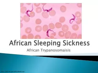
Introduction • Human African Trypanosomiasis (HAT) - sleeping sickness, is a major cause of mortality and morbidity in sub-Saharan Africa • Sleeping sickness, is a disease caused by infection with protozoan parasite and transmitted by the tsetse fly • Trypanosoma brucei gambiense (West African form) • Trypanosoma brucei rhodesiense (East African form) • Tsetse transmitted trypanosomiasis continues to be a major public health problem in districts bordering the Lake Victoria-River Nile- Lake Kyoga
Introduction • In the past decade, rhodesiense sleeping sickness has continued to be reported in: • North and Eastern Uganda in districts of: • Soroti, Kaberamaido and now Lira
Objectives • To assess the sleeping sickness status north of Lake Kyoga, Uganda • To establish the risk of gambiense and rhodesiense HAT overlap in Lango and Teso Sub regions
Materials and Methods • Study Area • Aloi is in lira district North of lat 2ºN • Lira is supplied by the river systems of Aswa and Pager
Materials and Methods • Methods used were to assess • prevalence of trypanosomiasis in the study areas • also to ascertain the possibility of overlap of T. b. rhodesiense and T. b. gambiense
Lymph node aspirate • All patients were systematically screened for enlarged cervical lymph nodes (CLN) • When CLN was enlarged, they were punctured: • The fresh aspirate is expelled onto a slide, • Cover slips were applied to spread the sample aspirate to facilitate the reading • The wet preps were then quickly examined by microscope (M ×40) for the presence of motile trypanosomes
CATT • Card agglutination test for trypanosomiasis (CATT) is a specific test for T.b.gambiense • All patients were screened with CATT in order to ascertain the possibility of T. b. gambiense in the known T. b. rhodesiense area
Wet film • Examination of wet film was done on all suspects • In wet blood films, 5 to 10 μl of finger prick blood was placed on a slide and examined microscopically (M ×40) under a cover slip
Heamatocrit Centrifugation Technique (HCT) • HCT was used to screen all the patients given the fact that it concentrates the parasites in one field for easy detection of the parasites • Capillary tubes containing anticoagulant were three-quarters filled with finger prick blood • The dry end sealed with plasticine • By high-speed centrifugation about 13,000 rpm for 6 to 8 min, trypanosomes are concentrated at the buffy coat between the plasma and the erythrocytes • The capillary tubes were mounted in a special holder, and directly examined at low magnification (×10 or ×16) for mobile parasites
Animal inoculation • Each isolate was inoculated in both Mastomys natalensis and Swiss mice with a view that: • if infection established in both species, then the infection is T.b. rhodesiense, • but if infection established only in Mastomys rats, then we suspect T.b. gambiense • parasitaemia examined after a period of 3 days
DNA analysis • Samples were analyzed using Loop Mediated Isothermal Amplification (LAMP) for Detection of HAT • DNA was extracted from parasites and mammalian cells using “GENE ALL” total DNA extraction kit for blood • LAMP was carried out with the Loopamp DNA amplification kit (Eiken Chemical Co. Ltd, Japan) • LAMP reaction mixture (25ul) contained template DNA, 40 pmol each of FIP and BIP (inner primers), 5 pmol each of F3 and B3 (outer primers), 8U of Bst DNA polymerase large fragment (New England Biolabs Inc.) • 1.4 mM deoxynucleoside triphosphates, 0.8 M betaine, 20 mM Tris-HCL (pH 8.8), 10 mM KCL, 10 mM (NH4)2SO4, 8 mM MgSO4, and 0.1% Tween 20 • A negative control reaction was incorporated by omitting template DNA from one reaction • The reaction mixture was incubated at 65ºC for 1 hr and heated at 80ºC for 2 min to terminate the reaction
Results: Rat inoculations • Parasitaemia was established in both Swiss mice and Mastomyssp. rats • No positive agglutination test was revealed by CATT, a test specific for T.b. gambiense
Disease stage • Eight of the cases taken to Lwala Hospital: • were stage II with WBC count > 5 cells/mm3 and trypanosomes in CSF. • only one was stage I with < 5 cells/mm3 and no CNS involvement.
Results: DNA analysis • Six of the 9 HCT detected cases were found positive by LAMP reaction for T. b. rhodesiense detection • Samples were also picked from 8 suspected patients who were HCT negative but had presenting signs like: • lymph node enlargement and • fever and • The suspected cases tested negative with lamp.
Discussions • A new sleeping sickness focus north of Lake Kyoga has been revealed in Aloi, Lira district • The trypanosome species isolated from the study area was T. b. rhodesiense • Infections established in both rodent species • No positive agglutination test was revealed by CATT, a test specific for T.b.gambiense. Thus, there is no T.b.gambiense overlap in this area • The exercise rules out the possibility of HAT overlap of • T.b. gambiense and T.b.rhodesiense within the study area so far
Acknowledgements • EANETT • DDHS: Lira, Soroti, Kaberamaido, Dokolo • Directorates of Production: Lira, Soroti, Kaberamaido, Dokolo • Director, NaLIRRI • Prof. Sugimoto (Hokkaido, Jp)