Download
1 / 35
370 likes | 516 Views
Respiratory distress. 1-tachypnea (RR>60/min) 2-retraction ( subcostal – intercostal – suprasternal ) ↑ WOB 3-grunting 4-flaring of ala nasi 5-cyanosis 6-rales and wheezing. Respiratory distress. Time of onset? Cyanosis response to oxygen? Chest X ray?. RDS risk factors. prematurity,

E N D
Respiratory distress 1-tachypnea (RR>60/min) 2-retraction ( subcostal – intercostal – suprasternal ) ↑ WOB 3-grunting 4-flaring of ala nasi 5-cyanosis 6-rales and wheezing
Respiratory distress Time of onset? Cyanosis response to oxygen? Chest X ray?
RDS risk factors prematurity, maternal diabetes perinatal asphyxia, twin pregnancy, cold stress, precipitous labor, cesarean delivery. The incidence is highest in preterm male or white infants.
Reduced risk of RDS The risk of RDS is reduced in pregnancies with: chronic or pregnancy-associated hypertension, maternal heroin use, prolonged rupture of membranes, and antenatal corticosteroid prophylaxis.
Pathophysiology lack of surfactant leads to progressive atelectasis, V/Q mismatch, hypoxia, hypercapnia, respiratory and metabolic (lactic) acidosis, pulmonary hypertension, right to left shunting through ductus arteriosus and foramen ovale.
Pathology the lungsof infants who die of RDS have a characteristic uniformly ruddy and airless appearance macroscopically resembling hepatic tissue. On microscopic examination the prominent finding is diffuse atelectasis and hyaline membrane.
Surfactant Therapy: Animal Models Expansion patterns in lung sections of a newborn rabbit: 1a - after treatment with natural surfactant 1b - no treatment
Radiographic Changes WithExogenous Surfactant Treatment before after
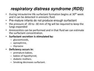
Surfactant is present inhigh concentrations in fetal lung homogenates by 20 wk of gestation,but it does not reach the surface of the lungs until later. • It appears in amniotic fluid between 28 and 32 wk. • Mature levelsof pulmonary surfactant are usually present after 35 wk. • Synthesis of surfactant depends in part on normal pH, temperature,and perfusion. • Asphyxia, hypoxemia, and pulmonary ischemia, particularly in association with hypovolemia, hypotension,and cold stress, may suppress surfactant synthesis.
Clinical manifestations infants with RDS present at birth or within several hours after birth with clinical signs of respiratory distress including tachypnea, retraction, grunting, nasal flaring and cyanosis. A late onset of tachypnea should suggest other conditions.
Clinical manifestations Some patients require resuscitation at birth because of intrapartum asphyxia or initial severe respiratory distress (especially with a birthweight <1,000 g). Cyanosis is relatively unresponsive to oxygen administeration.
Clinical manifestations The natural course of untreated RDS is characterizedby progressive worsening of cyanosis and dyspnea. If thecondition is inadequately treated, blood pressure may fall;fatigue, cyanosis, and pallor increase, and grunting decreases ordisappears as the condition worsens. Apnea and irregular respirationsoccur as infants tire and are ominous signs requiringimmediate intervention. Patients may also have a mixed respiratory-metabolic acidosis, edema, ileus, and oliguria. Respiratoryfailure may occur in infants with rapid progression of the disease.
Clinical manifestations In most cases, the symptoms and signs reach a peak within 3 days,after which improvement is gradual. Improvement is often heraldedby spontaneous diuresis and the ability to oxygenate theinfant at lower inspired oxygen levels or lower ventilator pressures. Death is rare on the 1st day of illness, usually occursbetween days 2 and 7, and is associated with alveolar air leaks(interstitial emphysema, pneumothorax), pulmonary hemorrhage,or IVH. Mortality may be delayed weeks or months if BPDdevelops in mechanically ventilated infants with severe RDS.
Chest X Ray the typical radiographic features (not pathognomonic) consist of a diffuse reticulogranular pattern giving the classic “ground-glass appearance” in both lung fields with superimposed airbronchograms and hypoaeration of the lung.
Differential diagnosis • in pneumonia manifested at birth, the chest roentgenogram may be identical to that for RDS. Maternal group B streptococcal colonization, organisms on Gram stain of gastric or tracheal aspirates or a buffy coat smear, and/or the presence of marked neutropenia may suggest the diagnosis of early-onset sepsis. • Cyanotic heart disease (total anomalous pulmonary venous return) can also mimic RDS both clinically and radiographically. Echocardiography with color flow imaging should be performed in infants who fail to respond to surfactant replacement to rule out cyanotic congenital heart disease as well as ascertain patency of the ductus arteriosus and assess pulmonary vascular resistance.
Differential diagnosis Persistent pulmonary hypertension, aspiration (meconium, amniotic fluid) syndromes, spontaneous pneumothorax, pleural effusions, and congenital anomalies such as cystic adenomatoid malformation, pulmonary lymphangiectasia, diaphragmatic hernia, and lobar emphysema must be considered, but can generally be differentiated from RDS by roentgenographic evaluation.
PREVENTION • Avoidance of unnecessary or poorly timed cesareansection, appropriate management of high-risk pregnancy andlabor, and prediction and possible in utero acceleration of pulmonaryimmaturity are important preventivestrategies. • In timing cesarean section or induction of labor, estimationof fetal head circumference by ultrasonography and determinationof the lecithin concentration in amniotic fluid by thelecithin:sphingomyelin ratio (particularly useful with phosphatidylglycerolin diabetic pregnancies) decrease the likelihoodof delivering a premature infant. • Antenatal and intrapartum fetalmonitoring may similarly decrease the risk of fetal asphyxia;asphyxia is associated withan increased incidence and severityof RDS.
PREVENTION Administration of betamethasone to women 48 hr before thedelivery of fetuses between 24 and 34 wk of gestation significantlyreduces the incidence, mortality, and morbidity of RDS. Corticosteroid administration is recommended for all women inpreterm labor (24-34 wk gestation) who are likely to deliver afetus within 1 wk.
PREVENTION Prenatal glucocorticoid therapy decreasesthe severity of RDS and reduces the incidence of other compiicationsof prematurity, such as IVH, patent ductus arteriosus(PDA), pneumothorax, and necrotizing enterocolitis, withoutadversely affecting postnatal growth, lung mechanics or development,or the incidence of infection. Prenatal glucocorticoidsmay act synergistically with postnatal exogenous surfactanttherapy.
PREVENTION Prenatal dexamethasone may be associated with ahigher incidence of periventricular leukomalacia than betamethasone. The relative risk of RDS, IVH and death is higher withantenatal dexamethasone treatment when compared withbetamethasone.. Administration of a 1st dose of surfactant into the trachea ofsymptomatic premature infants immediately after birth (prophylactic)or during the 1st few hours of life (early rescue) reducesair leak and mortality from RDS, but does not alter the incidenceof BPD.
Treatment The basic defect requiring treatment is inadequatepulmonary exchange of oxygen and carbon dioxide; metabolicacidosis and circulatory insufficiency are secondary manifestations.
Treatment For the 1st 24 hr, 10% glucoseand water should be infused through a peripheral vein at a rateof 65-75 ml/kg/24 hr. Excessive fluids (>140cc/kg/day) contribute to the development of PDA and BPD.
Goal ABG pao2=55-70, paco2=45-55 and pH=7.25- 7.45
Antibiotics should be started, Because pneumonia may present with the same clinical and radiological signs.
Dopamine should be started at 5-10mcg/kg/min to support circulation in the case of hypotension. Relief of metabolic and respiratory acidosis by bicarbonate and assisted ventilation may cause a fall in blood pressure.
Surfactant 1- 3 doses of intratracheal surfactant in the first 48 hr of life . The earlier the administration , the better the result.
Respiratory support The goal of respiratory support is establishment of appropriate oxygen saturation (90 -95%) and ABG . Different levels of respiratory care : 1- oxygen via oxyhood 2-oxygen via nasal pronge 3- nasal CPAP 4- intubation and mechanical ventilation
Nasal prong Oxyhood
Nasal CPAP Oxygen mask
Respiratory failure PH< 7.2 PaCO2> 60 mmHg PaO2< 50 mmHg or Frequent apnea Despite inspiring 100% O2.
Other causes of respiratory distress 1-pneumonia 2-meconium aspiration 3- transient tachypnea of newborn 4-TE fistula 5-diaphragmatic paralysis 6-vocal cord paralysis 7-congenital diaphragmatic hernia 8- cystic adenomatoid malformation 9-bronchogenic cyst 10-congenital lobar emphysema 11-congenital heart disease 12-pulmonary lymphangiectasis 13- pneumothorax 14-PPHN 15-choanal atresia 16-pulmonary hypoplasia 17-pulmonary sequestration