Download
1 / 31
310 likes | 352 Views
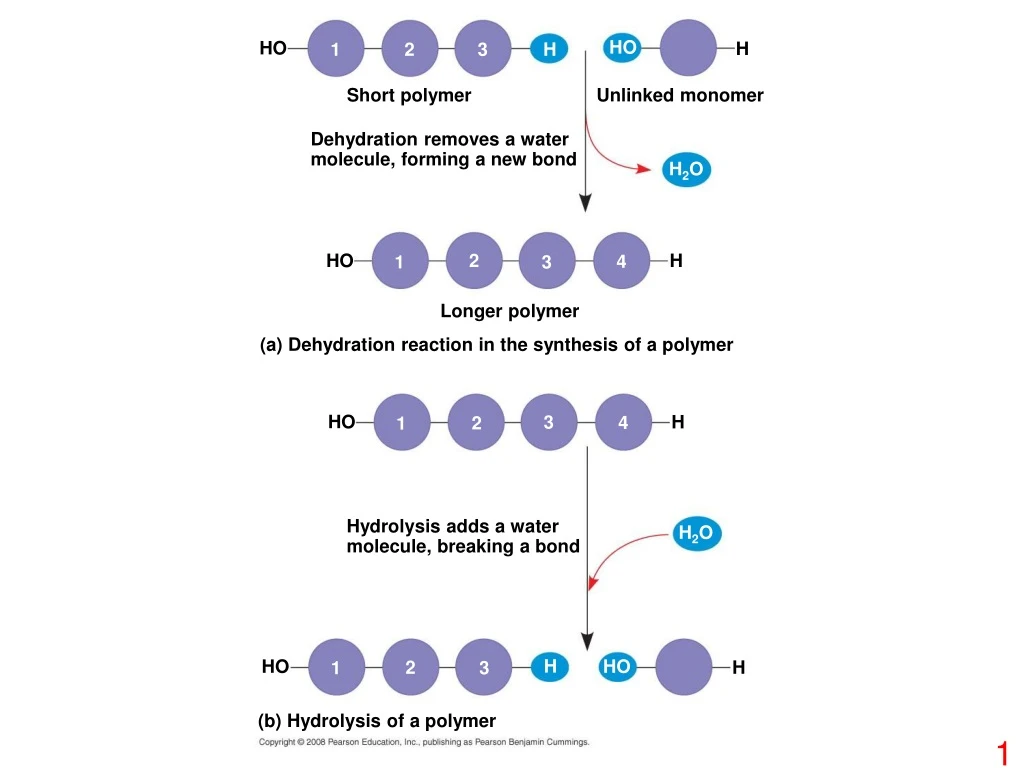
HO. HO. H. 1. 2. 3. H. Short polymer. Unlinked monomer. Dehydration removes a water molecule, forming a new bond. H 2 O. HO. 2. H. 4. 1. 3. Longer polymer. (a) Dehydration reaction in the synthesis of a polymer. HO. H. 3. 4. 2. 1. Hydrolysis adds a water

E N D
HO HO H 1 2 3 H Short polymer Unlinked monomer Dehydration removes a water molecule, forming a new bond H2O HO 2 H 4 1 3 Longer polymer (a) Dehydration reaction in the synthesis of a polymer HO H 3 4 2 1 Hydrolysis adds a water molecule, breaking a bond H2O H HO HO 2 H 1 3 (b) Hydrolysis of a polymer
Trioses (C3H6O3) Pentoses (C5H10O5) Hexoses (C6H12O6) Aldoses Glyceraldehyde Ribose Glucose Galactose Ketoses Dihydroxyacetone Ribulose Fructose
(a) Linear and ring forms (b) Abbreviated ring structure
1–4 glycosidic linkage Glucose Glucose Maltose (a) Dehydration reaction in the synthesis of maltose 1–2 glycosidic linkage Glucose Fructose Sucrose (b) Dehydration reaction in the synthesis of sucrose
Mitochondria Chloroplast Starch Glycogen granules 0.5 µm 1 µm Glycogen Amylose Amylopectin (a) Starch: a plant polysaccharide (b) Glycogen: an animal polysaccharide
(a) and glucose ring structures Glucose Glucose (b) Starch: 1–4 linkage of glucose monomers (b) Cellulose: 1–4 linkage of glucose monomers
Cell walls Cellulose microfibrils in a plant cell wall Microfibril 10 µm 0.5 µm Cellulose molecules • Glucose monomer
(a) (c) (b) Chitin forms the exoskeleton of arthropods. The structure of the chitin monomer. Chitin is used to make a strong and flexible surgical thread.
Fatty acid (palmitic acid) Glycerol (a) Dehydration reaction in the synthesis of a fat Ester linkage (b) Fat molecule (triacylglycerol)
Structural formula of a saturated fat molecule Stearic acid, a saturated fatty acid (a) Saturated fat Structural formula of an unsaturated fat molecule Oleic acid, an unsaturated fatty acid cis double bond causes bending (b) Unsaturated fat
Choline Phosphate Hydrophilic head Glycerol Fatty acids Hydrophobic tails Hydrophilic head Hydrophobic tails Space-filling model Phospholipid symbol (a) (b) (c) Structural formula
Hydrophilic head WATER Hydrophobic tail WATER
Substrate (sucrose) Glucose Enzyme (sucrase) OH H2O Fructose H O
carbon Carboxyl group Amino group
Nonpolar Glycine (Gly or G) Valine (Val or V) Leucine (Leu or L) Isoleucine (Ile or I) Alanine (Ala or A) Trypotphan (Trp or W) Methionine (Met or M) Phenylalanine (Phe or F) Proline (Pro or P) Polar Glutamine (Gln or Q) Serine (Ser or S) Threonine (Thr or T) Cysteine (Cys or C) Tyrosine (Tyr or Y) Asparagine (Asn or N) Electrically charged Acidic Basic Glutamic acid (Glu or E) Histidine (His or H) Aspartic acid (Asp or D) Lysine (Lys or K) Arginine (Arg or R)
Peptide bond (a) Side chains Peptide bond Backbone Amino end (N-terminus) Carboxyl end (C-terminus) (b)
Groove Groove (a) (b) A ribbon model of lysozyme A space-filling model of lysozyme
Protein from flu virus Antibody protein
Tertiary Structure Primary Structure Secondary Structure Quaternary Structure pleated sheet +H3N Amino end Examples of amino acid subunits helix
Hydrophobic interactions and van der Waals interactions Polypeptide backbone Hydrogen bond Disulfide bridge Ionic bond
Polypeptide chain Chains Iron Heme Chains Hemoglobin Collagen
Normal hemoglobin Sickle-cell hemoglobin Primary structure His Val Leu Glu Glu His Thr Val Primary structure Thr Pro Val Leu Pro Glu 1 2 3 4 5 6 7 1 2 3 4 5 6 7 Exposed hydrophobic region Secondary and tertiary structures Secondary and tertiary structures subunit subunit Sickle-cell hemoglobin Quaternary structure Normal hemoglobin (top view) Quaternary structure Function Molecules interact with one another and crystallize into a fiber; capacity to carry oxygen is greatly reduced. Function Molecules do not associate with one another; each carries oxygen. 10 µm 10 µm Red blood cell shape Normal red blood cells are full of individual hemoglobin moledules, each carrying oxygen. Red blood cell shape Fibers of abnormal hemoglobin deform red blood cell into sickle shape.
Denaturation Denatured protein Normal protein Renaturation
Correctly folded protein Polypeptide Cap Hollow cylinder Steps of Chaperonin Action: Chaperonin (fully assembled) The cap attaches, causing the cylinder to change shape in such a way that it creates a hydrophilic environment for the folding of the polypeptide. The cap comes off, and the properly folded protein is released. 2 3 1 An unfolded poly- peptide enters the cylinder from one end.
DNA 1 Synthesis of mRNA in the nucleus mRNA NUCLEUS CYTOPLASM mRNA 2 Movement of mRNA into cytoplasm via nuclear pore Ribosome 3 Synthesis of protein Amino acids Polypeptide
5 end Nitrogenous bases Pyrimidines 5C 3C Nucleoside Nitrogenous base Thymine (T, in DNA) Uracil (U, in RNA) Cytosine (C) Purines Phosphate group Sugar (pentose) 5C Adenine (A) Guanine (G) (b) Nucleotide 3C Sugars 3 end (a) Polynucleotide, or nucleic acid Ribose (in RNA) Deoxyribose (in DNA) (c) Nucleoside components: sugars
5' end 3' end Sugar-phosphate backbones Base pair (joined by hydrogen bonding) Old strands Nucleotide about to be added to a new strand 3' end 5' end New strands 5' end 3' end 5' end 3' end