Download
1 / 34
420 likes | 496 Views
Prolactin. Prolactin (PRL) is a single chain 198 amino acid polypeptide secreted by the anterior pituitary . Structurally similar to GH, its receptors resemble GH receptors , Prolactin also binds to specific receptors in the gonads, lymphoid cells, and liver Pulsatile , circadian fashion

E N D
Prolactin Prolactin (PRL) is a single chain 198 amino acid polypeptide secreted by the anterior pituitary . Structurally similar to GH, its receptors resemble GH receptors, Prolactin also binds to specific receptors in the gonads, lymphoid cells, and liver Pulsatile, circadian fashion Highest –sleep Nadir (lowest) – 10 am to noon Reference Range Serum PRL 1-25ng/mL or ug/L for females 1-20ng/mL or ug/L for males
Factors affecting PRL secretion Factors increasing PRL secretion Estrogen (during pregnancy stimulates lactotropes to secrete PRL) Breast feeding (reflex increase) TRH Stress sleep Dopamine antagonists chest wall stimulation or trauma Factors inhibiting PRL secretion Dopamine Bromocryptine, metoclopramid(Dopamine agonist) Somatostatin PRL (by negative feedback)
Prolactin action • PRL is responsible for Lactogenesis(initiation of lactation) & galactopoiesis(continuation of lactation) • - Stimulates milk production in the breasts by formation of Casein & Lactalbumin • Stimulates breast development along with Estrogen during pregnancy • Inhibits Ovulation by decreasing secretion of LH & FSH • May be involved in development of Leydig cells in pre-pubertal males • Immunomodulatory effects– stimulates T cell functions • Prolactin receptors in thymus
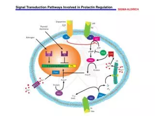
Regulation of Prolactin secretion • Secretion of prolactin is under tonic inhibitory control by dopamine, which acts via D2-type receptors located on lactotrophs • Prolactin production can be stimulated by the hypothalamic peptides, thyrotropin-releasing hormone (TRH) and vasoactive intestinal peptide (VIP) • positive feedback by sucking infant
Causes of Hyperprolactinemia • Hypothalamic Dopamine Deficiency • Diseases of the hypothalamus( including tumors, arterio-venous malformations, and inflammatory processes ) • Drugs (e.g. alpha-methyldopa and reserpine) • Defective Transport Mechanisms • Section of the pituitary stalk • Pituitary or stalk tumors • Lactotroph Insensitivity to Dopamine • Dopamine-receptor-blocking agents: phenothiazines (e.g. chlorpromazine), butyrophenones (haloperidol), and benzamides (metoclopramide, sulpiride, and domperidone) • Stimulation of Lactotrophs • Hypothyroidism- increased TRH production (acts as a PRF) • Estrogens: stimulate lactotrophs • Injury to the chest wall: abnormal stimulation of the reflex associated with the rise in prolactin that is seen normally in lactating women during suckling • Ectopic secretion of prolactin by nonpituitary tumors
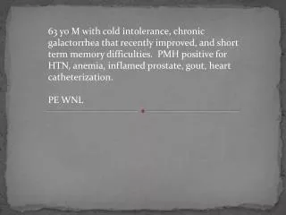
Clinical Features of Hyperprolactinemia/Prolactinoma • rule out secondary causes • Women • Oligomenorrhea • Amenorrhea (because GnRH release is decreased in direct response to PRL, inhibits release of FSH & LH ) • galactorrhea • infertility • Hirsutism • Menoften have less symptoms than women , Tend to present later - Gynaecomastia • Impotence-- often ignored • Most common functional pituitary tumor usually a microadenoma • space occupying macroadenoma • In both sexes, tumor mass effects may cause visual-field defects or headache • Treatment: Dopamine agonists like Bromocryptine which decreases PRL secretion
Prolactinoma • Diagnosis • Assess hypersecretion • Basal, fasting morning PRL levels (normally <20 ug/L) • Multiple measurements may be necessary • Pulsatile hormone secretion • levels vary widely in some individuals with hyperprolactinemia • Both false-positive and false-negative results may be encountered • May be falsely lowered with markedly elevated PRL levels (>1000 ug/L) • assay artifacts; sample dilution is required to measure these high values accurately • May be falsely elevated by aggregated forms of circulating PRL, which are biologically inactive (macroprolactinemia) • Hypothyroidism should be excluded by measuring TSH and T4 levels • Exclude Drugs which decrease dopamine stores • If prolactin level > 200, almost always a prolactinoma (even in a nursing mothers) • Prolactin levels correlate with tumor size in the macroadenomas • Suspect another tumor if prolactin low with a large tumor
ACTH • also known as corticotropin • ACTH is made up of 39 amino acids • is a polypeptide tropic hormone produced in the anterior pituitary by proteolytic processing of Prepro-opiomelanocortin (POMC). • Other neuropeptide products include b and g lipotropin, b-endorphin, and a-melanocyte-stimulating hormone (a-MSH) the role of it in anterior pituitary is not completely understood • ACTH principal effects are increased production and release of corticosteroids. • Overproduction of ACTH may accompany increased pigmentation due to a-MSH
ACTH synthesis ACTH Processing and cleavage of pro-opiomelanocortin (POMC)
- Vasopressin potentiates CRH at both hypothalamic and pituitary levels. • Circadian pattern of release • Highest levels of cortisol are in early AM following ACTH release • pituitary tumor is the cause of elevated ACTH this is known as Cushing's Disease . • Cushing’s syndrome is a rare condition that is the result of too much of the hormone cortisol in the body regardless of the cause.Such patients present with signs and symptoms of the excess cortisol (hypercortisolism) .It is caused by: • ACTH producing pitu tumors • cortisol-like medications (called glucocorticoids) • ACTH producing ectopic tumors • Cortisol producing adrenal glands • Primary adrenal insufficiency, also called Addison's disease, occurs when adrenal gland production of cortisol is chronically deficient, resulting in chronically elevated ACTH levels, Skin color will darken
Disorders of ACTH secretion • Increase secretion • - CRH • Adrenalectomy • Stress: • Hypoglycemia • Surgery & trauma • anesthesia • Infection & pyrogens • ADH • Psychiatric disturbances: • Anxiety, depression • adrenergic antagonists • Serotonin • Acetylcholine • Interleukines • Gastrointestinal peptides Decrease secretion Increase cortisol ACTH Somatostatin Opiod Enkephalins GABA
ACTH deficiency (within 2 weeks) symptoms of cortisol insufficiency are developed • - nausea, vomiting, anorexia, fatigue, weakness • hypoglycemia:caused by • increased sensitivity of tissues to insulin • decreased glycogen reserves • decreased gluconeogenesis) • in women, loss of body hair and decreased libido • due to decreased adrenal androgen production • - No features of mineralocorticoid deficiency • Aldosterone secretion is unaffected
excessive secretion of corticotrophin (ACTH) central form ofCushing syndrome (Cushing disease) Causes: - micro- or macroadenomas of adenohypophysis -hypothalamic disorders Pathophysiology: Chronichypercortisolismis the main disturbance of ACTH Symptoms and signs: • weight gain:- accumulation of adipose tissue in the trunk, facial, and cervical areas (truncal obesity, moon face, buffalo hump) • - weight gain from Na and water retention glucose intoleranceDM type 2 polyuria:osmotic polyuria due to glycosuria
The ACTH stimulation • test (also called Synacthen test) is a medical test to assess the functioning of the adrenal glands stress response by measuring the adrenal response to adrenocorticotropic hormone (ACTH) . • ACTH is a hormone produced in the anterior pituitary gland that stimulates the adrenal glands to releasecortisolandDehydroepiandrosterone(DHEAS). • During the test, a small amount of synthetic ACTH is injected, and the amount of cortisol, and sometimes aldosterone, the adrenals produce in response is measured. This test may cause mild to moderate side effects in some individuals. • This test is used to diagnose or exclude primary and secondary adrenal insufficiency, Addison's disease and related conditions. In addition to quantifying adrenal insufficiency, the test can distinguish whether the cause is adrenal (low cortisol and aldosterone production) or pituitary (low ACTH production)
Melanocyte-stimulating hormone(MSH) • MSH peptides derived by proteolytic cleavage of POMC • a-MSH has antipyretic and anti-inflammatory effects • Also inhibits CRH and LHRH secretion • Four MSH receptors identified • May inhibit feeding behavior • ACTH has MSH-like activity • However– MSH has NO ACTH like activity
LH, FSH • Secreted from cells in anterior pituitary . • Synthesis and secretion stimulated by GnRH– major effect on LH • LH, FSH aand bsubunits, Each subunit encoded by different gene • a subunit is identical for all 4 hormones(TSH and hCG) • bsubunit are unique and provide biological specificity. • Pulsatile pattern of secretion • LH pulses are biphasic (every 1 minute, then large pulse at 1 hour) • FSH pulses are uniphasic • Diurnal– LH/FSH more pronounced during puberty • Cyclic in females– ovarian cycle with LH surge at time of ovulation • Males are not cyclic, but constant pulses of LH cause pulses of testosterone to be produced
Pulsatile secretion of GnRH and LH • FSH secretion controlled by inhibin • Pulsatile secretion of GnRH and inhibin cause distinct patterns of LH and FSH secretion
Inhibin was initially isolated from gonadal fluids based on its ability to selectively inhibit FSH secretion from pituitary cells. • Inhibin is a heterodimer composed of an α -subunit and a β A- or β B-subunit to form inhibin A or inhibin B, both of which are secreted from the ovary. • Activin is a homodimer of inhibin β subunits with the capacity to stimulate the synthesis and secretion of FSH. • -Inhibin B also plays an important negative feedback role on FSH, independent of estradiol, during the menstrual cycle. • - it unlikely that ovarian activin plays an endocrine role in FSH regulation. However, activin may play an autocrine /paracrine role in the ovary, and it may also act locally in the pituitary to modulate FSH production.
HORMONAL INTEGRATION OF THE NORMAL MENSTRUAL CYCLE • The sequence of changes in reproductive function is coordinated through a series of feedback loops that alter pulsatile GnRH secretion, the pituitary response to GnRH, and the relative secretion of LH and FSH from the gonadotrope. • Negative feed-back • Activin is produced in both pituitary gonadotropes and folliculostellate cells and stimulates the synthesis and secretion of FSH. • Inhibins function as potent antagonists of activins through sequestration of the activin receptors. Although inhibin is expressed in the pituitary, gonadal inhibin is the principal source of feedback inhibition of FSH. • For the majority of the cycle, the reproductive system functions in a classic endocrine negative feedback mode. Estradiol and progesterone inhibit GnRH secretion, and the inhibins act at the pituitary to selectively inhibit FSH synthesis and secretion . • This negative feedback control of FSH is critical to development of the single mature oocyte in women. • Testosterone from Leydig cells– synthesis stimulated by LH, feedsback to inhibit GnRH production from hypothalamus and down-regulates GnRH receptors • Positive feedback • The exponential rise in estradiol results in positive feedback on the pituitary, leading to the generation of an LH surge (and a smaller FSH surge), that is essential for ovulation of a mature follicle.
Disorders of Puberty • PRECOCIOUS PUBERTY • - Traditionally has been defined as the development of secondary sexual characteristics before the age of 8 in girls - More recently, if breast development or pubic hair were present at <7 years of age for Caucasian girls or <6 years for African-American girls. • It is characterized by pulsatile LH secretion and an enhanced LH and FSH response to exogenous GnRH (two- to threefold stimulation) . • True precocity is marked by advancement in bone age of >2 SD . • In girls, centrally mediated precocious puberty is idiopathic in ~85% of cases; however, neurogenic causes must also be considered.
DELAYED PUBERTY • Is the absence of secondary sexual characteristics by age 13 in girls. The diagnostic considerations are very similar to those for primary amenorrhea • - Between 25 and 40% of delayed puberty in girls is of ovarian origin, with Turner syndrome constituting a majority of such patients. • Functional hypogonadotropichypogonadism encompasses diverse etiologies such as systemic illnesses, including celiac disease and chronic renal disease, and endocrinopathies, such as diabetes and hypothyroidism. • abnormalities in energy balance that result from exercise, dieting, and/or eating disorders. Together these reversible conditions account for ~25% of delayed puberty in girls. • Congenital hypogonadotropichypogonadism in girls or boys can be caused by mutations in several different genes or combinations of genes .Family studies suggest that genes identified in association with absent puberty may cause delayed puberty and that there may be a genetic susceptibility to environmental stresses such as diet and exercise. • neuroanatomic causes of delayed puberty are considerably less common in girls than in boys, it is always important to rule these out in the setting of hypogonadotropichypogonadism.
Thyroid stimulating hormone(TSH) • Is also known as thyrotropin, is secreted from cells in the anterior pituitary cells(thyrotrophs) when stimulated by TRH • Its basic sequence is glutamic acid-histidine-proline • It acts on its receptors on epithelial cells in the thyroid gland, and stimulates it to synthesize and release thyroid hormones T3 and T4. • Pattern of secretion is relatively steady • TSH is a glycoprotein hormone composed of two subunits which are non-covalently bound to one another. • The alpha subunit of TSH is also present in two other pituitary FSH and LH, in the placental hormone hCG. • The α subunit is thought to be the effector region responsible for stimulation of adenylate cyclase (involved the generation of cAMP) • Each of these hormones also has a unique beta subunit, which provides receptor specificity.
Feedback control of thyroid function Secretion of TRH, and hence, TSH, is inhibited by high blood levels of thyroid hormones in a classical negative feedback
TSH Excess Benign tumor of the pituitary (adenoma) Congenital hypothyroidism (cretinism) Primary hypothyroidism i.e. Hashimoto's thyroiditis pregnancy,& trophoblastic tumors : hCGshows some cross-reactivity to the TSH receptor and therefore can stimulate production of thyroid hormones Laboratory findings
TSH Deficiency Grave`s disease: Hyperthyroidism caused by circulating antibodies to the TSH receptor. Associated with diffuse goiter. Autoantibodies bind to TSH receptor and mimic the action of TSH itself leads to persistent stimulation of thyroid and elevated levels of thyroid hormones • Somatostatinis also produced by from the hypothalamus, inhibits the pituitary production of TSH • Primary hyperthyroidism i.e. Graves' disease • Secondary hypothyroidism or "central" hypothyroidism • TSH concentrations are measured as part of a thyroid function test • TSH is secreted throughout life but particularly reaches high levels during the periods of rapid growth and development • reference range for adults to be reduced to 0.4–2.5 µIU/mL • 0.4–7.0 µIU/mL at 14 months age