Download
1 / 23
370 likes | 1.2k Views
INTEGUMENTARY SYSTEM. CHAPTER 16: SKIN. FUNCTIONS OF THE SKIN. Protects the body –prevents loss of water, salt, heat & against invasion of pathogens & toxins Lubricates skin surface (with sebum, secreted from sebaceous glands) Maintains temperature

E N D
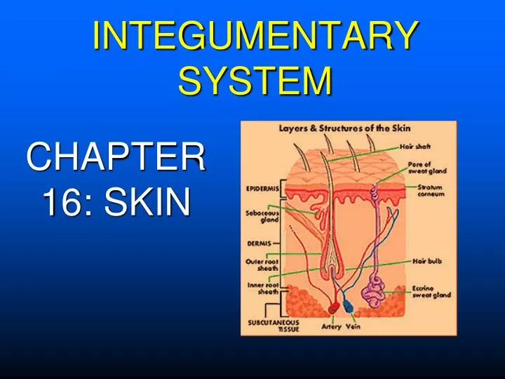
INTEGUMENTARY SYSTEM CHAPTER 16: SKIN
FUNCTIONS OF THE SKIN • Protects the body –prevents loss of water, salt, heat & against invasion of pathogens & toxins • Lubricates skin surface (with sebum, secreted from sebaceous glands) • Maintains temperature • Cools body by evaporation (with sweat secreted from sweat glands) • Has blood vessels that dilate & constrict • Has nerve fibers under the skin that aid in sensations of pain, pressure, touch, and temperature
LAYERS OF SKIN • Epidermis – outermost layer • Basal layer - deepest layer of epidermis • Stratum Corneum – cells are called “horny cells” or keratinized cells, outermost layer of epidermis • Lacks blood & lymph vessels & connective tissue • Corium (Dermis)– middle layer between epidermis and subcutaneous layer • Contains blood & lymph vessels, hair follicles, glands, and nerve fibers • Has connective tissue cells & elastic collagen fibers • Subcutaneous – innermost layer • Lipocytes (fat cells) mostly present; help to act as a heat insulator and for energy storage
Stratum corneum Basal layer melanocytes Epidermis Dermis Subcutaneous tissue You will need to be able to locate and/or identify individual layers of skin
ACCESSORY ORGANS OF SKIN • Hair – tight network of horny cells filled with keratin & melanocytes; keratin production determines hair color • Nails – hard keratin plates composed of horny cells • Glands • Sebaceous – located all over the body except palms of hands and soles of feet, produce sebum to lubricate skin • Sweat – coiled gland most numerous on palms of hands & soles of feet, sweat is a mixture of water and salt, functions to cool the body
Figure 16-2. (A) Anatomical structure of a nail. (B) Onycholysis. Infection or trauma to the nail may be the cause of the detachment of the nail from its plate. (B from Seidel HM: Mosby's Guide to Physical Examination, 5th ed. St. Louis, Mosby, 2003, p. 214.)
COMBINING FORMS RELATED TO SKIN diaphor / o = sweatdiaphor / esis = profuse sweating condition hidr / o = sweat an / hidr / osis = condition of not sweating ichthy / o = dry, scalyichthy / osis = dry, scaly skin myc / o = fungusdermat / o / myc / osis = fungal skin inf onych / o = nailonych / o / myc / osis =fungus in nail trich / o = hairtrich / o / myc / osis = fungus in hair xer / o = dryxer / o / derma = dry skin ungu / o = nail sub / ungu / al = under the nail adip / = fatadip / ose = pertaining to fat albin / o = whitealbin / ism = condition of white (skin) kerat / o = hard, horny tissue kerat / osis = abnormal condition of …… xanth / o = yellowxanth / oma = yellow mass (tumor) melan / o = blackmelan / oma = black tumor seb / o = oily (sebum)seb/ o / rrhea = oily discharge
TYPES OF SKIN LESIONS • Cyst – thick walled, closed sac or pouch containing fluid or semi-solid material • Fissure – groove or crack-like sore • Macule – discolored (often red) flat lesion
TYPES OF SKIN LESIONS • Papule – small solid elevation of the skin • Polyp – mushroom like growth extending on stalk from surface of mucous membrane • Pustule – small elevation in skin containing pus • Ulcer – open sore on skin or mucous membrane • Decubitus ulcer – bedsore, from staying in one position • Vesicle – small collection of clear fluid, blister • Wheal – smooth, slightly elevated swollen area that is more red or pale than surrounding skin (also called hive)
BULLAE Figure 16-4. Bullae (large blisters) in bullous pemphigoid (a chronic skin disorder in older individuals). The pemphigoid (pemphix means bubble) bullae occur as the entire thickness of the epidermis detaches from its foundation. (From Kumar V, Cotran RS, Robbins SL: Basic Pathology, 7th ed. Philadelphia, WB Saunders, 2003, p. 797.)
ABNORMAL SKIN SYMPTOMS • Alopecia – absence of hair from areas where hair normally grows • Pruritis – itching associated with many skin conditions • Urticaria – hives; acute allergic reaction where red, round wheals develop on skin • Figure 16-5. Dermatologic signs. • Alopecia areata. • (B) Ecchymoses, right hand. • (C) Petechiae. • (D) Senile purpura. Fragile blood vessels rupture with minimal trauma. • (E) Urticaria
ABNORMAL SKIN CONDITIONS • Impetigo – characterized by vesicles, pustules, and crusted over lesions; Rx – antibiotics and proper handwashing • Scabies – accompanied by severe itching; Rx – topical medications to destroy mites
ABNORMAL SKIN CONDITIONS • Tinea – named by location, Rx for all: antifungals • corpus – body, commonly called ringworm • tinea pedis – feet, commonly called athletes foot • tinea barbae – under a beard • tinea capitis – on the scalp Figure 16-9. (A) Tinea corporis (ringworm). (B) Tinea unguium. Fungal infection of the nail causes the distal nail plate to turn yellow or white. Hyperkeratotic debris accumulates, causing the nail to separate from the nail bed (onycholysis).
ABNORMAL SKIN CONDITIONS Wart – verruca is medical term for wart Figure 16-12. Verruca vulgaris. Warts are multiple papules with rough, pebble-like surfaces.
ACNE • Acne – papular and pustular lesions of the skin Figure 16-6. (A) Formation of a blackhead (comedo) in a dilated pore filled with sebum, bacteria, and pigment. (B) Acne vulgaris on the face.
ABNORMAL SKIN CONDITIONS • Eczema – inflammatory skin disease with red, pepulovesicular lesions • Psoriasis – chronic condition; itchy, scaly, red plaques on the skin covered by silvery grey scales Figure 16-8. Psoriasis. Scaly erythematous plaque, with silvery scales on top.
ABNORMAL SKIN CONDITONS Figure 16-10. Vitiligo on the hand (Latin: vitium meaning a 'blemish'). Epidermal melanocytes are completely lost in depigmented areas through an autoimmune process.
Figure 16-11. (A) Callus on the sole of the foot. (B) Keloid. (A from Mosby's Medical, Nursing, and Allied Health Dictionary, 6th ed., St. Louis, Mosby, 2002, p. 265; B from Ignatavicius DD, Workman ML: Medical-Surgical Nursing: Critical Thinking for Collaborative Care, 4th ed. Philadelphia, WB Saunders, 2002, p. 1544.)
ABNORMAL SKIN CONDITIONS • Cellulitis – acute infection of skin marked by heat, redness, pain, and swelling; Rx: antibiotics • Gangrene – death of tissue from loss of blood supply
BURNS • First degree – superficial burn, affecting epidermal layer of skin, skin is red and dry; Ex – sunburn • Second degree – affecting epidermal and dermal layers; skin is red and has blisters; Ex - burn from cooking • Third degree – epidermal and dermal layers destroyed, subcutaneous tissue damaged, charred white, grey and/or black tissue Figure 16-7. Burns. (A) Second-degree injury. Wound sensation is painful and very sensitive to touch and air currents. (B) Third-degree burn showing viable color (deep-red, white, black and brown). The wound itself is insensate (does not respond to pinprick sensation).
ABNORMAL SKIN CONDITIONS • Actinic Keratosis – precancerous skin lesion (usually caused by sun exposure) • Squamous Cell Carcinoma -malignant tumor of squamous epithelial cell of epidermis, remember epithelial cells also line internal organs so you can have squamous cell carcinoma in the mouth, larynx, lung, etc… Figure 16-15. (A) Actinic (solar) keratosis. (B) Squamous cell carcinoma. Lesions are often nodular and ulcerated.
ABNORMAL SKIN CONDTIONS • Basal Cell Carcinoma – malignant tumor of basal cell layer of epidermis • Karposi’s Sarcoma - tumors develop in the tissues below the skin surface, or in the mucous membranes. Common in HIV+ patients Figure 16-13. (A) Basal cell carcinoma. (B) Kaposi sarcoma.
ABNORMAL SKIN CONDITIONS • The ABCDs of melanoma. • Asymmetry: one half unlike the other half. • Border: irregular or poorly circumscribed border. • Color: varied from one area to another; shades of tan & brown; black; sometimes white, red or blue. • Diameter: usually larger than 6mm (diameter of a pencil eraser).