Download
1 / 54
540 likes | 745 Views
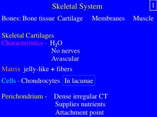
Skeletal System. 1. Bones: . Bone tissue. Cartilage . Membranes . Muscle . Skeletal Cartilages. Characteristics - . H 2 O. No nerves. Avascular . Matrix. jelly-like + fibers. Cells -. Chondrocytes. In lacunae. Perichondrium -. Dense irregular CT. Supplies nutrients.

E N D
Skeletal System 1 Bones: Bone tissue Cartilage Membranes Muscle Skeletal Cartilages Characteristics - H2O No nerves Avascular Matrix jelly-like + fibers Cells - Chondrocytes In lacunae Perichondrium - Dense irregular CT Supplies nutrients Attachment point
Skeletal Cartilages 2 Hyaline - Support with flexibility & resilience Articular Nose Costal Respiratory passages Elastic - Withstand repeated bending External ear Epiglottis Fibrocartilage - Prevent compressive shock Intervertebral discs Menisci Pubic symphysis
Hyaline Cartilage Outer ear Tip of nose Articulations (Joints) * Costal Intervertebral disc * * * * * * Pubic symphysis Elastic Cartilage Trachea Epiglottis Fibrocartilage Menisci 3
Bone Functions 4 1. Support - Hard framework Attachment points 2. Protection - Vital organs 3. Movement - Levers for muscles 4. Mineral homeostasis - Ca+2 & PO4-3 5. Nutrient & growth factor storage - Yellow marrow Insulin-like & transforming growth factors Bone morphogenic
6. Hematopoiesis 5 Red marrow Blood cell formation Redmarrow In infants: All medullary cavities Spongy bone In adults: Head of the femur & humerus Vertebrae Diploë of flat bones Coxal bone Severe anemia: Yellow marrow
Bone Texture http://trc.ucdavis.edu/mjguinan/apc100/modules/Musculoskeletal/bone/structure0/structure.html Compact bone - Dense outer surface Osteon Spongy bone - Inner layer Trabeculae – small needlelike Red or yellow marrow Epiphyseal line 6
Typical Long Bone 7 Compact bone Yellow marrow Red marrow Diaphysis - Tubular shaft Periosteum Endosteum Medullary cavity - Adipose tissue Epiphyses - Expanded Interior is spongy bone Exterior is compact bone Articular cartilage Epiphyseal line - Hyaline cartilage Remnant epiphyseal plate
8 Red marrow Articular cartilage Epiphysis Epiphyseal line Endosteum Medullary cavity Yellow marrow Diaphysis Periosteum Epiphysis Nutrient artery Spongy bone Compact bone
9 Epiphysis Articular cartilage Diaphysis Spongy bone Hyaline Compact bone Periosteum http://www.xecu.net/kiirenza/anatomy/pictures/longbone1.jpg
10 Medullary cavity Red marrow Endosteum http://www.xecu.net/kiirenza/anatomy/chicken.htm
Short, irregular & flat bones 11 Spongy Outside: Thin plates Compact Periosteum Inside: Diploë Endosteum Marrow RedorYellow
Chemical Composition of Bone 12 Sacrificial fractures Organic components – 30% 1. Cells - Osteoclasts, osteoblasts, osteocytes, osteogenic 2. Osteoid - Ground substance - Proteoglycans & glycoproteins Fibers - Collagen Function: Flexibility & tensile strength Resist stretch & twist Resilience
Inorganic Components 13 Collagen fibers 50 – 65% by mass Hydroxyapatites (mineral salts) Calcium carbonate Calcium phosphate Tiny crystals Function: Hardness Resist compression
1. Osteogenic Cell division 2. Osteoblast 1 2 3 4 3. Osteocyte 4. Osteoclast Stem cell from mesenchyme Secretes bone matrix Ruffled border Maintains daily metabolism Resorb bone 14
Osteoblast 15 Osteocyte Osteocyte http://www3.umdnj.edu/histsweb/lab4/bone/bonecells.html Osteoclasts
Osteoclast 16 http://neuromedia.neurobio.ucla.edu/campbell/bone/wp_images/89%20osteoclast.jpg Resorption 50 monocytes Digestive enzymes & acids Maintain blood Ca+2 levels
Compact bone 15 17 Vessels & nerves Lamellae - Lamellae Blood vessels & nerves Structural & functional unit: Osteon Haversian system Parallel pillars Concentric hollow tubes Haversian canal - Central tube Function: Resist torsion
Compact Bone 18 Periosteum Central canals Medullary cavity Lacuna Cytoplasmic arms Volkman’s canals Perforating canals Transverse Function: Osteocyte Canaliculi Gap junctions Function: Homeostasis Transport Communication
Microscopic compact bone 19 Osteocyte Haversian canal Canaliculi Lamellae Osteon Medullary cavity Spongy bone Volkman’s canal Periosteal artery Periosteum Osteon or Haversian system
20 Osteon (Haversian System) Haversian canal
21 Canaliculi Osteocytes in lacunae Haversian canal
Spongy Bone 22 Red marrow No osteons Trabeculae Trabecular cavities Short, flat (diploe) & irregular Epiphysis Function: Support & protect red bone marrow
Spongy Bone 23 Trabeculae Osteocyte Endosteum Osteoclast Osteoblasts Red marrow
Membranes and Blood Supply 24 Periosteum - Exterior, white Fibrous layer - Dense irregular CT Nerves, lymphatic & blood vessels Periosteal arteries Volkmann’s Nutrient artery Nutrient foramen Osteogenic - Bone forming Osteoblasts & osteoclasts Function: Increase diameter Insertion points Fracture repair Nourishment Endosteum - Delicate CT Trabeculae Lines canals Osteoblasts & osteoclasts
Bone Tissue Formation 25 Fibrous CT & hyaline cartilage Skull & clavicle Osteoblast Osteoid Periosteum Woven bone Compact bone Red marrow Broken down Replaced Ossification = Osteogenesis @ week 8 Intramembranous Ossification Fibrous CT membranes Mesenchymal Diploe Endochondral ossification - Hyaline cartilage
Endochondral Ossification - Embryonic 26 Perichondrium Periosteum Chondrocytes die Nutrient Artery 2. Cavitation 3. Periosteal bud 1. Bony collar Model Calcify Hyaline Osteoclasts – erode cartilage Blood vessels Osteoblasts Osteoblasts - Spongy bone
Birth 27 Arteries Articular cartilage Secondary ossification Epiphyseal Plate 5. Epiphyses ossify 4. Medullary cavity Osteoclasts break down Bones formed: All Osteoblasts - compact Except skull & clavicle
Bone Growth in Length 28 Epiphyseal plate Hyaline cartilage Chondrocytes proliferate, mature & die Cartilage matrix calcifies Osteoclasts dissolve calcified cartilage Osteoblasts secrete bone matrix
Epiphyseal Plate 29 http://www.rit.edu/~mrppph/animals/pages/epiphyseal_plate.htm
Epiphyseal Plate Epiphysis 30 Diaphysis Resting Zone Anchors plate to bone Growth Zone Cell division Pushes epiphysis Hypertrophic Zone Chondrocytes enlarge Matrix calcifies Osteogenic Zone Osteoclasts Osteoblasts
Factors Affecting Bone Growth 31 Osteocytes Minerals Ca & P F, Mg, Fe, Mn Vitamins C: Collagen Osteoblasts K & B12 Proteins – alkaline phosphatase A Stimulates Osteoblasts Hormones
Childhood 32 Ends bone growth Pituitary - hGH Function: Stimulate cell division & protein synthesis Thyroid – T3 & T4 Function: modulate growth to proportion Puberty Sex steroids – estrogen & androgens Adrenal glands, adipose tissue Function: Adolescent growth spurt Testosterone masculinizes Estrogen feminizes
Bone Remodeling - Adults 33 Osteoclasts & Osteoblasts Periosteum & Endosteum Bone deposit - Osteoblast Secrets an osteoid seam - Hydroxyapatite crystals Function: Repair bone injury Add extra strength Bone resorption - Osteoclast Lysosomal enzymes, acids Ca+2 salts soluble Function: Blood Ca+2 homeostasis
Ca+2 Homeostasis 34 9-11 mg/100 ml Intestines 99% in bone Nerve cells Cofactor Coagulation Contraction Stimulus - Low Ca+2 Receptor & control center - Parathyroid glands Output - Parathyroid hormone (PTH) Effector - Osteoclasts stimulated to resorb bone Kidneys secrete calcitriol Response - Releases Ca+2 to the blood
Stimulus - 35 Electrical signals 1% per week Mechanical stress High Ca+2 Receptor & control center - Thyroid gland Output - Calcitonin (CT) Effector - Osteoblasts stimulate Ca+2 salt deposits Osteoclasts are inhibited Response - Ca+2 removed from blood Mechanical stressors - Muscle pull Gravity Stimulates remodeling Strengthens the skeleton Hormones = when Stress = where
Fractures 36 Position of the bones Nondisplaced - Displaced - Completeness Complete - Incomplete - Orientation relative to long axis Linear - Transverse - Penetration of skin Open (compound) - Closed (simple) -
37 Complete transverse fracture that is displaced
Repair simple fracture Periosteal Haversian canal Inflammation 1. Hematoma formation Blood clot Bone cells die Macrophages 38
2. Fibrocartilage callus 39 Fibroblasts Chondroblasts Capillaries Granulation tissue Osteogenic stem cells Fibrocartilage 3 weeks
3. Bony callus formation 40 Osteoblasts Osteogenic stem cells Spongy bone 2 - 3 months
4. Bone remodeling 41 Remove excess bone Compact bone Osteoclasts Osteoblasts 3-4 months
Primary Germ Layers Ectoderm Mesoderm Endoderm http://www.med.unc.edu/embryo_images/unit-mslimb/mslimb_htms/mslimb009.htm 16 days 42
43 Neural groove http://www.med.unc.edu/embryo_images/unit-mslimb/mslimb_htms/mslimb009.htm 22 days
25 Day Embryo 44 http://www.med.unc.edu/embryo_images/unit-mslimb/mslimb_htms/mslimb009.htm
Notochord 27 days 45 http://www.med.unc.edu/embryo_images/unit-mslimb/mslimb_htms/mslimb009.htm Directs formation of skeleton
Skeleton Development 46 Leg Arm Leg Arm http://evolution.berkeley.edu/evosite/evo101/IIC1Homologies2.shtml Limb buds @ week 4
Limb Development 47 http://www.med.unc.edu/embryo_images/unit-mslimb/mslimb_htms/mslimb009.htm 64 days http://www.med.unc.edu/embryo_images/unit-mslimb/mslimb_htms/mslimb009.htm 51 days
Bone Disorders 49 Osteomalacia - Soft bones Osteoid produced; no Ca+2 salts deposited Cause: Ca+2 or Vitamin D Rickets Osteoporosis - Brittle bones Bone resorption> bone deposit Cause: Declining estrogen or testosterone levels Insufficient exercise Ca+2, protein, Vitamin D Smoking - Reduces estrogen Hyperparathyroidism
Osteomalacia 50


