Download
1 / 43
430 likes | 609 Views
X-Ray Rounds Plain Chest Radiographs. Garry W. K. Ho, M.D. VCU / Fairfax Family Practice July 13, 2005. The 12-Step Program. }. 1 : Name 2 : Date 3 : Old films 4 : What type of view(s) 5 : Penetration 6 : Inspiration 7 : Rotation 8 : Angulation

E N D
X-Ray RoundsPlain Chest Radiographs Garry W. K. Ho, M.D. VCU / Fairfax Family Practice July 13, 2005
The 12-Step Program } • 1: Name • 2: Date • 3: Old films • 4: What type of view(s) • 5: Penetration • 6: Inspiration • 7: Rotation • 8: Angulation • 9: Soft tissues / bony structures • 10: Mediastinum • 11: Diaphragms • 12: Lung Fields Pre-read } Quality Control } Findings
Pre-Reading • 1. Check the name • 2. Check the date • 3. Obtain old films if available • 4. Which view(s) do you have? • PA / AP, lateral, decubitus, AP lordotic
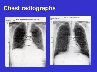
Quality Control • 5. Penetration • Should see ribs through the heart • Barely see the spine through the heart • Should see pulmonary vessels nearly to the edges of the lungs
Overpenetrated Film • Lung fields darker than normal—may obscure subtle pathologies • See spine well beyond the diaphragms • Inadequate lung detail
Underpenetrated Film • Hemidiaphragms are obscured • Pulmonary markings more prominent than they actually are
Quality Control • 6. Inspiration • Should be able to count 9-10 posterior ribs • Heart shadow should not be hidden by the diaphragm 1 2 3 4 5 6 7 8 9 10
Poor inspiration can crowd lung markings producing pseudo-airspace disease 8 9 About 8 posterior ribs are showing 9-10 posterior ribs are showing With better inspiration, the “disease process” at the lung bases has cleared
Quality Control • 7. Rotation • Medial ends of bilateral clavicles are equidistant from the midline or vertebral bodies
If spinous process appears closer to the right clavicle (red arrow), the patient is rotated toward their own left side If spinous process appears closer to the left clavicle (red arrow), the patient is rotated toward their own right side
Quality Control • 8.Angulation • Clavicle should lay over 3rd rib 1 2 3
Apical lordotic Same patient, not lordotic Pitfall Due to Angulation A film which is apical lordotic (beam is angled up toward head) will have an unusually shaped heart and the usually sharp border of the left hemidiaphragm will be absent
Findings • 9. Soft tissue and bony structures • Check for • Symmetry • Deformities • Fractures • Masses • Calcifications • Lytic lesions
Findings • 10. Mediastinum • Check for • Cardiomegaly • Mediastinal and Hilar contours for increase densities or deformities
Findings • 11. Diaphragms • Check sharpness of borders • Right is normally higher than left • Check for free air, gastric bubble, pleural effusions
Findings • 12. The Lung Fields! • To help you determine abnormalities and their location… • Use silhouettes of other thoracic structures • Use fissures
Silhouette / Structure Contact with Lung Upper right heart border/ascending aorta Anterior segment of RUL Right heart border RML (medial) Upper left heart border Anterior segment of LUL Left heart border Lingula (anterior) Aortic knob Apical portion of LUL (posterior) Anterior hemidiaphragms Lower lobes (anterior) Lung Fields: Using Structures / Silhouettes
Lung Fields: Using Structures / Silhouettes Upper right heart border / ascending aorta (anterior RUL) Aortic knob (Apical portion of LUL ) Upper left heart border (anterior LUL) Right heart border (medial RML) Left heart border (lingula; anterior) Anterior hemidiaphragms (anterior lower lobes)
Major Oblique Fissure Separates the LUL from the LLL Right Major Fissure Separates the RUL/RML from the RLL Right Minor Fissure Separates the RUL from the RML Lung Fields: Fissures • The fissures can also help you to determine the boundaries of pathology
Now for the Cases… Remember… be systematic!
PA view: RML consolidation and loss of right heart silhouette Lateral View: RML wedge shaped consolidation RML pneumonia
RUL infiltrate / consolidation, bordered by minor fissure inferiorly Patchy LLL infiltrate that obscures the left hemidiaphragm; right and left heart borders obscured RUL and LLL pneumonia
Underpenetrated; possible nonspecific obscuring of left heart border; mostly normal
Multiple bilateral cavitary lesions with air-fluid levels c/w pulmonary abscesses Tuberculosis
28 y/o female with sudden onset SOB while jogging this morning Well demarcated paucity of pulmonary vascular markings in right apex Left spontaneous pneumothorax
RML consolidation that appears wedge shaped on lateral view RML pneumonia
RLL infiltrate / consolidation RLL pneumonia
Patient BIBA to ER s/p airplane crash. Widened mediastinum Concern for aortic injury
Explain the prominence of the right atrium on this AP radiograph Patient rotated to their right (left shoulder forward)
Dilatation of the main pulmonary artery with decreased peripheral vascular markings ?? Pulmonary embolism ??
Obscuring of the right and left heart borders; infiltrate at the bases Bilateral aspiration pneumonia
Diffuse bilateral fluffy interstitial infiltrates Pneumocystis carinii pneumonia
Left lung opacity Later diagnosed as lung cancer
Cardiomegaly, increased pulmonary vascular markings, fluid in the horizontal fissure CHF
What do the arrows indicate? Kerley B Lines Short (1 -2 cm) white lines at the lung bases, perpendicular to the pleural surface representing distended interlobular septa
The 12-Step Program } • 1: Name • 2: Date • 3: Old films • 4: What type of view(s) • 5: Penetration • 6: Inspiration • 7: Rotation • 8: Angulation • 9: Soft tissues / bony structures • 10: Mediastinum • 11: Diaphragms • 12: Lung Fields Pre-read } Quality Control } Findings