Download
1 / 52
750 likes | 2.56k Views
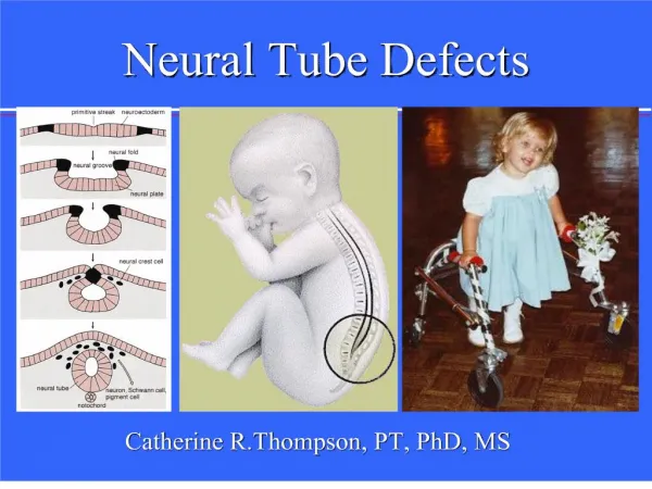
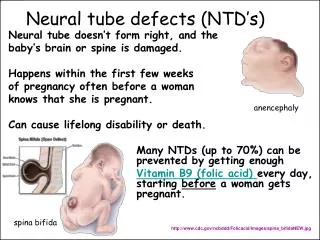
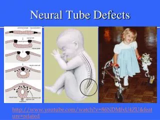
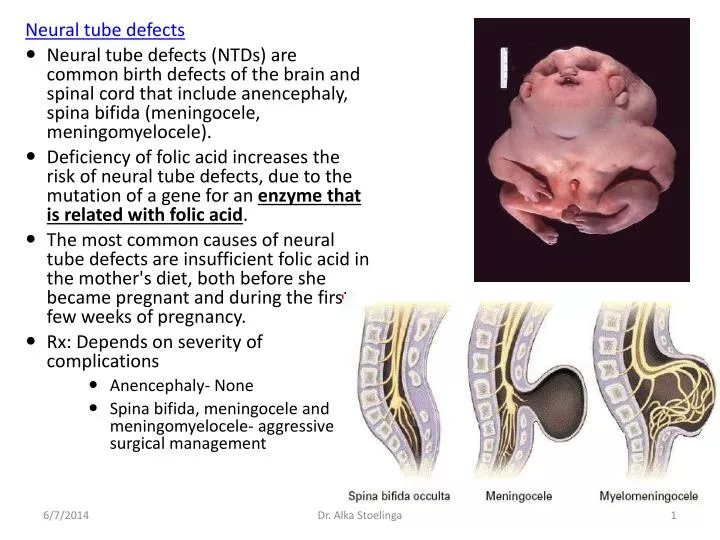
Neural tube defects Neural tube defects (NTDs) are common birth defects of the brain and spinal cord that include anencephaly, spina bifida (meningocele, meningomyelocele).

E N D
Neural tube defects • Neural tube defects (NTDs) are common birth defects of the brain and spinal cord that include anencephaly, spina bifida (meningocele, meningomyelocele). • Deficiency of folic acid increases the risk of neural tube defects, due to the mutation of a gene for an enzyme that is related with folic acid. • The most common causes of neural tube defects are insufficient folic acid in the mother's diet, both before she became pregnant and during the first few weeks of pregnancy. • Rx: Depends on severity of complications • Anencephaly- None • Spina bifida, meningocele and meningomyelocele- aggressive surgical management Dr. AlkaStoelinga
BROWN SEQUARD SYNDROME • Results from unilateral cord compression/ lateral hemisection of spinal cord • Aka- crossed hemiplegia • Impaired pain and temperature sensation • Impaired light touch and vibration and position sensation • Increased tendon reflexes and extensor plantar response Dr. Alka Stoelinga
Brown-Séquard syndrome's symptoms: * = Side of the lesion • Hypertonic paralysis • Spastic paralysis and loss of vibration and proprioception (position sense) and fine touch • Loss of pain and temperature sensation Dr. Alka Stoelinga
The hemisection of the cord results in a lesion of each of the three main neural systems: • the principal upper motor neuron pathway of the corticospinal tract • one or both dorsal columns • the spinothalamic tract • As a result of the injury to these three main brain pathways the patient will present with three lesions: • The corticospinal lesion produces spastic paralysis on the same side of the body (the loss of moderation by the UMN). • The lesion to fasciculus gracilis or fasciculus cuneatus results in ipsilateral loss of vibration and proprioception (position sense) as well as loss of all sensation of fine touch. • The loss of the spinothalamic tract leads to pain and temperature sensation being lost from the contralateral side beginning one or two segments below the lesion. Dr. Alka Stoelinga
CENTRAL CORD SYNDROME: • Involves gray matter and crossing of Spinothalamic tract • Motor weakness • Dissociate sensory loss • E.g:Syringiomyelia,Tumors ANTERIOR 2/3rd SYNDROME: • Bilateral involvement of anterior spinal cord • Motor,sensory,Autonomic functions are lost • Posterior column spared • E.g: Vascular:Thrombosis of anterior spinal artery or by tumors Dr. Alka Stoelinga
Patterns of sensory loss Dr. Alka Stoelinga
NON COMPRESSIVE SPINAL CORD LESION • Vascular • Inflammatory • Transverse myelitis • Multiple Sclerosis 3.Development:Syringomyelia 4.Metabolic: Subacute combined degeneration(Deficiency of Vit B12) Dr. Alka Stoelinga
Clinical differentiation: Dr. Alka Stoelinga
CERVICAL SPONDYLOSIS Dr. Alka Stoelinga
Spondylosis : • Spondylosis is a term referring to degenerative osteoarthritis of the joints between the centra of the spinal vertebrae and/or neural foraminae • When the space between two adjacent vertebrae narrows, compression of a nerve root emerging from the spinal cord may result in radiculopathy • sensory and motor disturbances, such as severe pain in the neck, shoulder, arm, back, and/or leg, accompanied by muscle weakness. • Less commonly, direct pressure on the spinal cord (typically in the cervical spine) may result in myelopathy • characterized by global weakness, gait dysfunction, loss of balance, and loss of bowel and/or bladder control. • The patient may experience a phenomenon of shocks (paresthesia) in hands and legs because of nerve compression and lack of blood flow. • If vertebrae of the neck are involved it is labeled as Cervical Spondylosis. • Lower back Spondylosis is labeled Lumbar Spondylosis. Dr. Alka Stoelinga
Physical signs of compression of cervical roots Dr. Alka Stoelinga
Investigations • Plain X ray cervical spine Antero-posterior Lateral • In severe cases MRI of the cervical spine • Management of cervical radiculopathy • Analgesic • Cervical collar • Surgery if deficit is severe or conservative therapy fails. Dr. Alka Stoelinga
Cervical collar Dr. Alka Stoelinga
Cervical spondylotic myelopathy : • Progressive, gradual onset • Prone to have hyperextension injury of the cervical cord • Spastic quadriparesis with loss of sensation and ultimately, involvement of bowel and bladder. • Investigation of choice is MRI • Treatments • conservative • surgery disc resection, vertebral lamina resection.Surgery sometimes leads to acute deterioration. Dr. Alka Stoelinga
Lumbar disc herniation : • Common problem in middle aged and elderly. • Precipitated by trauma or lifting a heavy weights when spine is flexed. • Onset may be sudden or gradual, Constant aching pain in the lumbar region and may radiate to the buttock, thigh, calf and foot. • Pain is exacerbated by coughing or straining and may be relieved by lying flat. • SLRT (straight leg rising test) may be positive (positive Lasegue’s sign) • MRI is the investigation of choice • Management :conservative or surgery Dr. Alka Stoelinga
Case • A 25 year old male was brought to Emergency department. Status- Post motor vehicle accident. • Emergency doctor does ABC management • On CNS examination • Motor • B/L Lower extremity weakness ~4/5 • B/L With hyperreflexia • Cranial nerves • Intact • Sensory • Pain and temperature- lost in lower extremities • Vibration and position- intact • Light touch- intact • What is the likely diagnosis? Why? Dr. Alka Stoelinga
Syringomyelia • Cavitation of spinal cord • Fluid-filled cavity (or cavities) develops near the centre of the spinal cord, usually in the cervical segments • The expanding cavity disrupts second-order spinothalamic neurons • May extend laterally to damage the anterior horn cells, and may compress the long fiber tracts. • It is assumed that the disturbed CSF dynamics cause the development of the syrinx but the mechanism is not clear. Dr. Alka Stoelinga
Communicating Associated with Arnold Chiari malformation • Noncommunicating secondary to spinal cord trauma Dr. Alka Stoelinga
Syringomyelia Dr. Alka Stoelinga
Clinical features • Pain in the neck or shoulder is common and patients may seek advice because of sensory loss in the upper limbs • Dissociated sensory loss (impaired pain and temperature sensation with preservation of dorsal column modalities- intact sensation of light touch) • Loss of protective sensory function leads to tropic lesions such as painless burns or ulcers on the hands • Ultimately sensory loss in all four limbs with UMN signs below the lesion and LMN sign at the level of lesion. • Investigation is MRI • Treatment is surgical. Dr. Alka Stoelinga
Subacute combined degeneration • Occurs with vitamin B12 deficiency • Distal paresthesias • Weakness of extremities • Spastic paresis • Ataxia • In classical case Deficit of vibration and proprioception with pyramidal signs (Plantar extension and hyperreflexia) • Investigation Serum Vitamin B12 level (Low) • Rx Vitamin B12 therapy • 250 µg to 1 mg of B12 daily • S/C or I/M injections of Vit B12 weekly for ~20 weeks followed by lifelong Dr. Alka Stoelinga
Anterior Spinal artery infarct • Acute flaccid paralysis which evolves into flaccid paresis over days to weeks • Loss of pain and temperature sensation • Sparing of vibration and position sense • (Posterior columns are supplied by Posterior spinal artery) Dr. Alka Stoelinga
Transverse myelitis:- • It affects one to five persons per million. • Transverse myelitis is an acute inflammatory condition usually secondary to viral illness or recent vaccination may be with multiple sclerosis and other inflammatory and vascular disorders (eg:-syphilis) • where there is a progressive sensory loss and weakness. • Transverse myelitis (TM) is an uncommon neurological syndrome caused by inflammation (includes swelling, pain, heat, and redness) of the spinal cord. • Characterized by weakness, back pain, and bowel and bladder dysfunction. Dr. Alka Stoelinga
Clinical feature: • Though non compressive, it presents as compressive myelopathy • Features depend on spinal segment involved • Commonly involved: Thoracic segments • Acute/ Subacute course • Paresthesia • Motor weakness: Paraplegia, UMN type • Sensory loss • Bladder involvement • Acute stage: Neural shock • May be difficult to differentiate with GBS • Similar symptoms may be present in multiple sclerosis Dr. Alka Stoelinga
Investigations: • MRI of spinal cord • Treatment: 1. Steroid: Methylprednisolone IV for 3 days Followed by Prednisolone 1mg/Kg/Day for several weeks 2. Other Rx: • Physiotherapy • Care of Bladder and bowel • Prevention of DVT Dr. Alka Stoelinga
Poliomyelitis: • Poliomyelitis results from a relatively selective destruction of lower motor neurons in the anterior horn cell of spinal cord by polio virus. • The disease causes Flaccid paralysis of muscles with accompanying Hyporeflexia and Hypotonicity. • Some patients may recover most function,whereas others progress to muscle atrophy and permanent disability. Dr. Alka Stoelinga
PARTIAL SEIZURES Simple partial seizure : • Not associated with loss of consciousness and Limited to part of the body • Denotes focal pathology in brain/involve only one hemisphere • Typically associated with structural abnormalities of brain such as scars,tumors,AV malformation or focal areas of inflammation • Can be Motor/Sensory/Autonomic/Psychomotor • In typical motor type there is clonic ( repetitive flexion and extension) movement at the rate of 2-3 Hz. • Other features of partial motor seizure are • Jacksonian March • Todd’s paralysis • Epilepsia partialis continua Dr. Alka Stoelinga
Partial sensory seizures-- somatic sensation like paraesthesia or tingling sensation /electric sensation in the contralateral face and limbs • Partial visual seizures—visual hallucinations such as ball of light,flashes of light and hallucinations of faces and scenes • Partial psychic seizures— • Sensation of falling /vertigo • There may be olfactory or auditory hallucinations, undue familiarity (déjà vu), feeling of unreality (jamais vu) • Autonomic seizures-- flushing, sweating, pilorection, epigastric discomfort, nausea Dr. Alka Stoelinga
Complex partial seizures: --Usually arises from temporal lobe and less frequently from frontal lobe Psychomotor(Temporal lobe) seizures: -- Associated with altered consciousness --Associated with loss of posture and tone --Aura may be present --Patients stop what he /she is doing and stares blankly, often making rhythmic smacking movements of lips/picking at their clothes --After few minutes patients gains consciousness but may be drowsy Dr. Alka Stoelinga
GENERALIZED SIEZURES • Involving diffuse regions of both hemisphere,simultaneously and synchronously • Results from cellular,biochemical or structural abnormalities with widespread distribution Absence seizures : • Petitmal epilepsy • Brief/Transient • Commonly seen in childhood • During an attack child stops activity • Vacant stares, may blink or roll up the eye and fails to respond to commands • Do not cause loss of posture • Attacks lasts for few seconds • More frequent Dr. Alka Stoelinga
Tonic seizures: Tonic tightening of limbs along with loss of consciousness • Clonic seizures: Clonic movements of limbs i.e rhythmic flexion and extension of limbs • Myoclonic seizures: It consists of single or multiple myoclonic jerks involving one part of the body or entire body. • Atonic seizures: Characterized by sudden loss of postural muscle tone,lasting for 1-2 secs,with brief impairment of consciousness Dr. Alka Stoelinga
Generalized tonic-clonic seizure : • Prodromal phase: Hours or days before attack,unease,irritability • Aura: Preceded by partial seizure,and patients usually anticipates that seizure will occur.These feeling may be olfactory hallucination,epigastricdiscomfort,jerking of one limb • Tonic phase: There is tonic contractions of the muscles,there is flexion and adduction of arms and extension of legs, patient goes rigid,presence of cyanosis and loss of consciousness. Lasts for 10-30 sec • Clonic phase: This period does not last for more than 1 min.There is violent jerky movements of face and limbs. Patient may sustain injuries like tongue bite and incontinence. Dr. Alka Stoelinga
Post-ictal phase : Unresponsiveness/Deep unconsciousness, muscular flaccidity, salivation, loss of corneal reflex,extensor plantar response.Lasts from few minutes to several hours • Patient gradually regains consciousness over minutes to hours. • There may be headache,vomiting,confusion,drowsiness,fatigue, muscle ache • Recover within 1-2 days • In EEG tonic phase has low voltage fast activity followed by high voltage polyspike discharges, spike and wave. • In post-ictal phase there is diffuse slowing Dr. Alka Stoelinga
D/D of seizure • TIA • Panic attacks • Syncope • Cardiac dysrhythmias • Metabolic ,Sleep disorders,Movement disorders Dr. Alka Stoelinga
Pseudo seizures: • Used to denote Hysterical conversion reactions • Attacks mimic epileptic seizures • Characterized by an asynchronous thrashing of the limbs,which increases if restraints are imposed. • There is no post ictal phase • EEG is normal • Increase prolactin level after 15-30 mints of true tonic-clonic seizure while unchanged in Pseudoseizure Dr. Alka Stoelinga
Anti epileptic drugs ( Anticonvulsant) • Phenytoin • Membrane stabilizing agent (↓Na and Ca influx) • Dose 3-6 mg/Kg/d • Adverse effects: gum hypertrophy, hirsutism, glucose intolerance, megaloblastic anemia, lymphadenopathy, nausea and vomiting, ataxia, nystagmus. • Teratogenic (fetal hydantoin syndrome) • Carbamazepine • Membrane stabilizing agent (↓Na influx) • Dose 15-35 mg /Kg/d • Adverse effects: ataxia, dizziness, diplopia, vertigo, aplastic anemia, leukopenia, hepatotoxicity. • Safe in pregnancy Dr. Alka Stoelinga
Phenobarbitone • MOA exactly unknown.may be involves in potentiation of GABA. • Dose 1-4 mg/Kg/d • Adverse effects: sedation, ataxia, nystagmus, vertigo, agitation , confusion, skin rash. • Valproic acid • Potentiation of GABA. • Dose 20-60 mg/Kg/bid-qid • Adverse effects: ataxia, sedation, tremor, hepatotoxicity, alopecia • Drug of choice in myoclonic seizure Dr. Alka Stoelinga
Ethosuximide • Reduces propagation of abnormal electrical activity in the brain • Dose 20-40 mg /Kg/ daily to bid • Ataxia, lethargy, headache, skin rash, GI irritation,bone marrow suppression • Drug of choice for absence seizures • Benzodiazepines • Clonazepam is effective in myoclonic and absence seizures. • Diazepam- immediate treatment of any seizure. • Good safety profile.Sedation, drowsiness occurs in high dose. Dr. Alka Stoelinga
Gabapentin and Lamotrigine • Gabapentin • is an analogue of GABA. • Effective in focal onset seizures • Side effects are sedation, dizziness, ataxia, GI irritation. • Dose 900-2500/d • Lamotrigine • inhibits exitatory neurotransmitters like glutamate and aspartate, blocks sodium channels. • Broad spectrum, dose is 150-500mg/d BID • Dizziness, diplopia, sedation, ataxia are adverse effects. Dr. Alka Stoelinga
EEG (Electro-Encephalogram) • An Electroencephalogram (EEG) is a test commonly performed to look at the electrical activity of the brain i.e. the brain waves or how the brain is functioning. • It is a simple, painless test and involves no needles or injections. • In normal subjects, 4 types of waves • In ascending order of frequency • δ.less than 4 waves per second • θ.4-6 waves per second • α.7-13 waves per second • β.more than 13 waves per second Dr. Alka Stoelinga
EEG cap Dr. Alka Stoelinga
EEG (Electro-Encephalogram) Delta waves Theta waves Location:Found in locations not related to task at hand focal subcortical lesions metabolic encephalopathy deep midline disorders some instances of hydrocephalus • Location:frontally in adults, posteriorly in children; high amplitude waves • subcortical lesions • diffuse lesions • metabolic encephalopathy hydrocephalus • deep midline lesions Dr. Alka Stoelinga
EEG (Electro-Encephalogram) Alpha waves Beta waves Location: both sides, symmetrical distribution, most evident frontally; low amplitude waves benzodiazepines • Location: posterior regions of head, both sides, higher in amplitude on dominant side. • coma Dr. Alka Stoelinga
EEG (Electro-Encephalogram) Gamma waves • Location:Somatosensory cortex • A decrease in gamma band activity may be associated with cognitive decline, especially when related the theta band Dr. Alka Stoelinga
Normal EEG Dr. Alka Stoelinga
Grossly abnormal EEG • Spikes and waves • Slowing of waves, appearance of delta wave • Triphasic slow waves • Electrocerebral silence Dr. Alka Stoelinga
Spikes and waves Dr. Alka Stoelinga