Download
1 / 47
500 likes | 1.32k Views
Chromosomal Abnormalities II SDK November 3, 2012. II- STRUCTURAL CHROMOSOMAL ABNORMALITIES. II- STRUCTURAL ABNORMALITIES. Theses are group of problems that is caused by the structural abnormalities of the chromosomes. Structural abnormalities may be Balanced or unbalanced

E N D
II- STRUCTURAL ABNORMALITIES • Theses are group of problems that is caused by the structural abnormalities of the chromosomes. • Structural abnormalities may be Balanced or unbalanced • Balanced. Where there is no loss or gain of chromosome material • Un Balanced. An `unbalanced’ translocation means that an individual has more or less chromosomal material than usual.(Loss or gain of genetic materail) • Structural abnormalities may occure in Germ cell or Somatic cells. • Those that happen in germ line such as cause difficulties in egg or sperm development and normal development of a zygote. This can be transferred to next generations • Those happen in Somatic cells can cause Cancer but are not transferred to next generation.
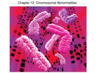
Types of Structural Abnormalities • Translocation • Reciprocal transloations • Robertsonian Translocation • Deletion and microdeletion • Duplication • Inversion • Pericentric inversion • Paracentricinversio • Insertion • Ring chromosome • Isochromosome • Chromosomal breakage/Instability: (e.g. Fanconi anemia, Bloom syndrome)
Translocation • A fragment of a chromosome is moved ("trans-located") from one chromosome to another - joins a non-homologous chromosome. Translocation are of 2 types • Reciprocal translocations • Robertsonian translocations
Examples of Reciprocal Abnormalities • Reciprocal Translocation of Chromosome 9 and 22(Produces Philadelphia Chromosome). This is because that the break occurred at an important gene which is actually an oncogene called “abl” oncogene this leads to CML(Chronic Myeloid Leukemia) • Reciprocal Translocation between Chromosome 8 and 14 result in overproduction of “myc” oncogene this leads to Burkitts Lymphoma.
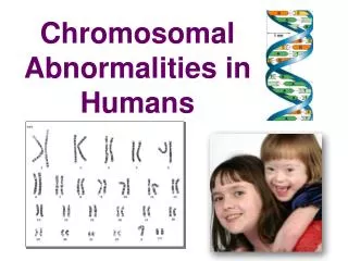
Down Syndrome • Traditional type(95%) that is trisomy 21 an extra 21 chromosome due to non disjunction. • Robertsonian Translocation Trisomy 21(5%). that is an extra q arm of chromosome 21 on chromosome 14. • In this case one of the parent may have Robertsonian Translocation and this is the cause of repeated abortions and miscarriage(Pregnancy loss). • So a female with a down child and repeated abortions might have Robertsonian Translocation
Terminal Deletion/ Cri Du Chat • Cri Du Chat: A specific Terminal deletion of a small portion of “chromosome 5” • These children have severe mental retardation, a small head with unusual facial features, and a cry that sounds like a distressed cat.
Interstitial Deletion • Prader-Willi and Angelman Syndrome • Deletion at chromosome 15.
Angelman, Prader-Willi syndromes • Usually caused by large (megabase+) interstitial deletions of 15q11-q13 • Delete maternal chromosome = AS • Delete paternal chromosome = PWS
Symptoms of Angelman Syndrome • Developmental delay • Functionally severe Speech impairment • Movement or balance disorder • Behavioral uniqueness: any combination of frequent laughter/smiling; apparent happy demeanor; easily excitable personality, often with hand flapping movements • Short attention span
What is Prader-Willi Syndrome • Prader-Willi syndrome is caused by the absence of normally active genetic material on the long arm of chromosome 15. • Deletion on the paternal chromosome 15 • Prevalence: 1:12,000- 15,000 (both sexes, all races)
Symptoms of Prader-Willi Syndrome • Poor weight gain in infancy • Excessive or rapid weight gain between 1 and 6 • Delayed sexual maturity • Mild to moderate mental retardation • Obsession with food
Duplication/ Fragile X Syndrome • If the fragment joins the homologous chromosome, then that region is repeated • Example Fragile X: one of the most common form of mental retardation. • The X chromosome of some people is unusually fragile at one tip - seen "hanging by a thread" under a microscope.
Duplication/ Fragile X Syndrome • Moderate to sever mental retardation • Speech delay, short attention, hyperactivity • Poor motor coordination and mouthing objects • Poor socialization, temper tantrum • Mood disorder (bipolar), schizophrenia
Types of inversion • Two types • Peri-centric. When inversion include the centromere is called peri-centric inversion. • Para-centric. When inversion do not include the centromere is called para-centric inversion.
Peri-centric Inversion of chromosome 16 & Small Partial trisomy of 16q. • A male infant born with full term pregnancy has • Hypospadiasis • Ambiguous genitalia • Poor sucking reflex • Poor growth • Microcephaly • Wide set eyes and depressed nasal bridge. • This baby father was suffering from Peri-centric inversion of chromosome 16 that leads to his son with double material at 16q causing small partial trisomy.
Ring chromosome • A ring chromosome is a chromosome whose arms have fused together to form a ring. • Ring chromosomes may form in cells following genetic damage by mutagens like radiation, they may also arise spontaneously during development.
Isochromosome An isochromosome is a chromosome that has lost one of its arms and replaced it with an exact copy of the other arm.
In a Robertsonian translocation fusion occurs at the: • Telomeres. • Centromeres. • Histones. • Ends of the long arms.
When karyotyping is needed • Dysmorphic features and/or developmental delay • Fetal / neonatal death, with multiple congenital malformations or dysmorphic features. • Indeterminate gender or ambiguous genitalia, amenorrhoea, infertility etc. • Recurrent miscarriages - if a couple have had 3 or more miscarriages - both members of the couple should be tested. • Known or suspected family history of chromosome abnormality (e.g. Down syndrome or Edwards syndrome), where the karyotype of the affected individual is not known or not available. • Known familial chromosome rearrangements e.g. Robertsonian or reciprocal translocations. • Terminated fetus, for confirmation of an abnormal cytogenetic result diagnosed previously.
Indications for fetal karyotyping • Raised maternal/ paternal age. • Ultrasound marker(s) indicative of chromosome abnormality • Previous fetus/child with chromosome abnormality. • Parent is known to carry a chromosome rearrangement. • Positive maternal serum or ultrasound screening for Down syndrome.
How to do Chromosome Analysis • A common example is chromosome analysis of amniotic fluid. • Amniotic fluid is the developing baby’s urine and trophoblastic secretions and therefore contains cells from the baby. • Chromosome analysis can also be performed on blood, skin cells, and other tissues.
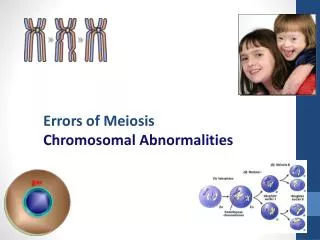
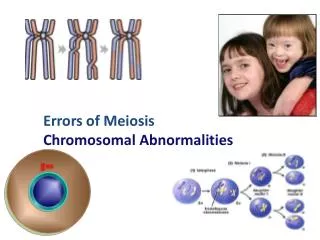
Summary • In each human cell, except the egg and sperm cells, there are 46 paired chromosomes of varying size • • One chromosome of each pair is inherited from each parent • • The autosomes are chromosomes numbered 1-22 (largest to smallest) • • The two sex chromosomes are called X and Y • • Egg cells contain 23 chromosomes, made up of 22 autosomes and an X • • Sperm cells contain 23 chromosomes, made up of 22 autosomes and an X or a Y • • When the egg and sperm join at conception, the baby will have 46 chromosomes in its cells, just like the parents • • Changes in the number, size or structure of chromosomes in the cells of an individual may cause a chromosomal condition that affects growth, development and health
Summary • A particular type of chromosomal structural change is called a translocation. There are two different types of translocations: • Reciprocal translocation - material is exchanged between any of the chromosomes and involves pieces of any size • Robertsonian translocation - material is exchanged between chromosomes 13, 14, 15, 21 and 22 • Where there does not appear to have been any loss or gain of chromosome material, the translocation is described as balanced • An unbalanced translocation means that an individual has more or less chromosomal material than usual