Download
1 / 16
200 likes | 1.04k Views
Molluscum Contagiosum. Yazid 17576. Molluscum Contagiosum. A self limited cutaneous infection caused by a large DNA poxvirus that affects both children and adults. DNA poxvirus, the largest virus known (200 X 300 X 100 nm), causes molluscum contagiosum (4 subtypes ).

E N D
MolluscumContagiosum Yazid 17576
MolluscumContagiosum • A self limited cutaneousinfection caused by a large DNA poxvirus that affects both children and adults. • DNA poxvirus, the largest virus known (200 X 300 X 100 nm), causes molluscumcontagiosum (4 subtypes). • Can be transmitted by direct skin contact, autoinoculation and vertical transmission. • The virus replicates in the cytoplasm of epithelial cells producing cytoplasmic inclusions, and it may cause enlargement of infected cells.
History • Molluscumcontagiosum appears to have a bimodal age distribution. • Childhood, (nonsexual skin contact). • Early adulthood (age 15-29 y), (sexually transmitted disease). • Most patients are asymptomatic; • some complains of pruritus, tenderness, and pain
History • The incubation is 14-50 days. • If patients have eczema or other diseases altering skin barrier function, molluscum may spread more rapidly in affected areas.
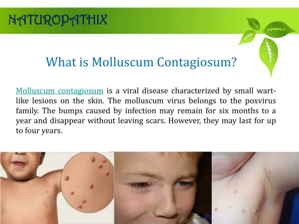
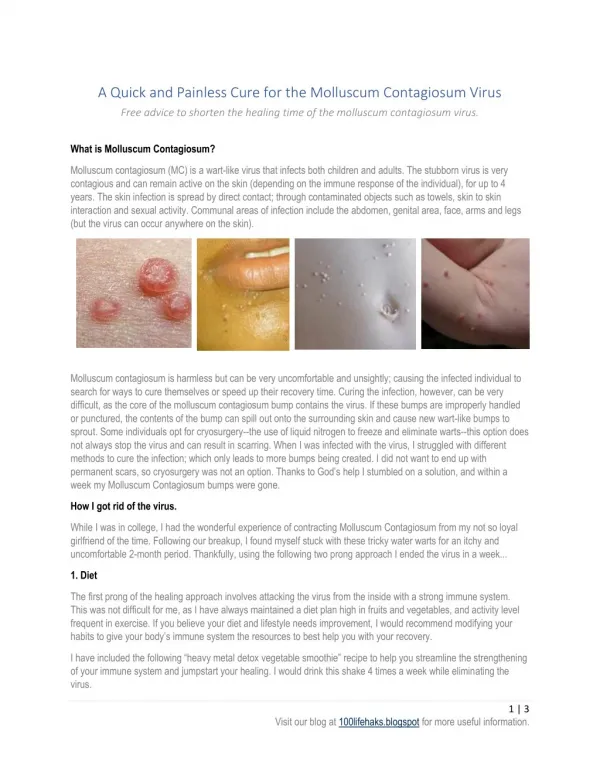
Signs • Skin - Primary lesion of molluscumcontagiosum • Firm, smooth, umbilicated papules, usually 2-6 mm in diameter (range 1-15 mm), may be present in groups or may be widely disseminated on the skin and mucosal surfaces. • The lesions can be flesh-colored, white, translucent, or even yellow in color. • The number of lesions varies from 1-20 up to hundreds in some reports. • Some lesions become confluent to form a plaque. • Lesions generally are self-limited but can persist for several years.
Signs Smooth, multiple, Umbilicated papules
Signs Molluscumcontagiosum may arise in areas that have been injured, often because they've been scratched. The papules form a row; this is known as koebnerisedmolluscum.
Signs Molluscumcontagiosum with Eczema and crusted lesions
Signs • The lesions can be flesh-colored, white, translucent, or even yellow in color. • Some lesions become confluent to form a plaque.
Signs • Skin - Distribution of molluscumcontagiosum • In children, papules are located mainly on the trunk and extremities. • In adults, lesions often are located on the lower abdominal wall, inner thighs, pubic area, and genitalia. • Although rarely found in the mouth or on the palms and soles, cases of molluscumcontagiosum involving the oral mucosa, including the lips, buccal mucosa, hard palate, retromolar pad, and tongue, have been reported.
Signs • Immunocompromisedconditions • multiple widespread, persistent, and disfiguring lesions can occur, especially on the face and possibly involving the neck and trunk.
DIFFERENTIAL DIAGNOSIS • Flat warts (HPV infection) • Large Solitary MolluscumKeratoacanthoma: squamous cell carcinoma, basal cell carcinoma, epidermal inclusion cyst. • Multiple Facial Mollusca in HIV-Infected Individual: Disseminated invasive fungal infection, i.e., cryptococcosis, histoplasmosis, coccidioidomycosis, penicillinosis.
LABORATORY EXAMINATIONS • Microscopic examination Giemsa-stained central semisolid core reveals “molluscum bodies” (inclusion bodies).
Treatment • Topical patient-directed therapy- 5% imiquimod cream applied at bedtime 3–5 times per week for up to 1–3 months. • Clinician-directed therapy - • Curettage - small mollusca can be removed with a small curette with little discomfort or pain. • Cryosurgery Freezing - lesions for 10–15 s is effective and minimally painful • Electrodesiccation - For mollusca refractory to cryosurgery, especially in HIV-infected individuals with numerous and/or large lesions
Complications & Prognosis • Complications of molluscumcontagiosum include irritation, inflammation, and secondary infections. • Lesions on eyelids may be associated with follicular or papillary conjunctivitis. • Molluscumcontagiosum is a benign, self-limited disease. • Treatments for molluscumcontagiosum are effective if patients are compliant. • Additional duration of therapy may be required in immunocompromised patients. • Overall, molluscumcontagiosum prognosis is excellent.