Download
1 / 26
270 likes | 290 Views
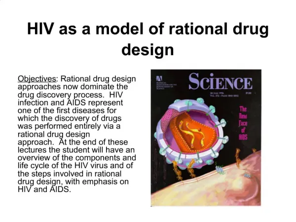
Rational Drug Design. Using the 3D Shape of Proteins to Design Drugs that Inhibit Protein Function. Before you start this activity, make sure you have the program Cn3D installed on your computer. Download Cn3d from this site. Examples of Protein Function. Hormones

E N D
Rational Drug Design Using the 3D Shape of Proteins to Design Drugs that Inhibit Protein Function Before you start this activity, make sure you have the program Cn3D installed on your computer. Download Cn3d from this site
Examples of Protein Function Hormones Insulin binds to receptors on cell membranes signalling cells to take up glucose from the blood Protein ChannelsRegulate movement of substances across the plasma membrane. E.g. The CFTR protein pumps ions across membranes Transport Haemoglobin (far right) in red blood cells transports oxygen to cells around the body Source: http://www.biology.arizona.edu/biochemistry/tutorials/chemistry/page2.html http://www.cbp.pitt.edu/bradbury/projects.htm http://www.abc.net.au/cgi-bin/common/printfriendly.pl?/science/news/enviro/EnviroRepublish_1191825.htm http://www.umass.edu/microbio/chime/
Catalase - enzyme power! Hydrogen peroxide, a natural product of metabolism in your cells, is highly toxic in high concentrations and must be removed quickly! Products Reactants oxygen Add ferric ions (Fe 3+) Rate increases 30 000-fold 2 2 water Hydrogen peroxide Add Catalase Rate increases 100 000 000-fold Location of active site where Hydrogen peroxide binds Source: http://accad.osu.edu/~ibutterf/ibp/molecule/ http://folding.stanford.edu/education/water.htm http://www.opti-balance.com/hyperox.htm
How enzymes do it! • Enzyme proteins have specific sites where all the action happens. We call this the active site. Molecules that need to be ripped apart or put together enter the active site. • Each protein has a specific shape so it will only perform a specific job. Ripping things apart Joining things together http://chsweb.lr.k12.nj.us/mstanley/outlines/enzymesap/Enzymesap.html http://academic.brooklyn.cuny.edu/biology/bio4fv/page/active_.html
Many toxins are proteins Ricin from the seeds of the castor oil plant destroys ribosomes Funnel web spider toxin: blocks movement of calcium ions. Source: http://www.wiley.com/legacy/college/boyer/0470003790/cutting_edge/molecular_recognition/molecular_recognition.htm http://science-univers.qc.ca/image/ricin061.jpg http://www.staabstudios.com/Spider.htm
Protein molecules are polymers • Proteins are very large polymer molecules. Polymers are made by linking smaller molecules, monomers, together to make a long chain. • In the case of proteins, the monomers are amino acids. There are 20 different amino acids. AA AA AA AA AA AA AA
Why is protein structure important? • Each protein molecule has a characteristic 3D shape that results from coiling and folding of the polymer chain. • The function of a protein depends upon the shape of the molecule.
Protein chains Each protein has a specific sequence of amino acids that are linked together, forming a polypeptide http://www.mywiseowl.com/articles/Image:Protein-primary-structure.png
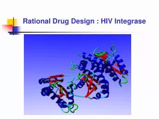
The protein chain folds Interactions between amino acids in the chain form: • alpha helices • beta sheets • Random coils Together usually form the binding and active sites of proteins Source: http://www.rothamsted.bbsrc.ac.uk/notebook/courses/guide/prot.htm#I
And folds again! • After folding, amino acids that were distant can become close • Now the protein chain has a 3D shape that is required for it to function correctly Source: io.uwinnipeg.ca/~simmons/ cm1503/proteins.htm
The final protein… The final protein may be made up of more than one polypeptide chain. The polypeptide chains may be the same type or different types. Source: http://fig.cox.miami.edu/~cmallery/150/chemistry/hemoglobin.jpg
Designing a Drug to Block Amylase Action Amylase is a protein that cuts small maltose sugar molecules off starch molecules. Another enzyme, maltase, is responsible for breaking down the maltose molecules into two simple sugars known as glucose. Glucose is absorbed into the blood and transported to cells around the body to provide them with energy. STARCH AMYLASE MALTASE GLUCOSE MALTOSE STARCH GLUCOSE
Block the active site of amylase Active Site Pig
Your turn…Designing a diet pill Click on the button on the right to start exploring amylase with its active site blocked by a drug. Amylase in Cn3D
Influenza Pandemics The Spanish Flu in 1918, killed approximately 50 million people. It was caused by the H1N1 strain of influenza A. The Asian Flu in 1957 was the H2N2 influenza A strain. Worldwide it is estimated that at least one million people died from this virus. The Hong Kong Flu in 1968 evolved into H3N2. 750,000 people died of the virus worldwide
Influenza epidemics • Economic Effects: • Days away from work • Providing medical advise and treatment • Mortalities Figure 1. Weekly number of influenza and pneumonia deaths per 10 000 000 population in the United States, France, and Australia (black line).
Designing a Flu Drug Step 1: looking for protein targets Influenza viruses are named according to the proteins sticking out of their virus coat. (H) There are two types of protein = Nand H. N and H have special shapes to perform specific jobs for the virus. (N)
N cuts the links between the viruses and the cell surface so virus particles are free to go and infect more cells. H attaches to cell surface proteins so virus can enter cell Virus Proteins on cell surface Virus genes are released into the cell. The lung cell is ‘tricked’ into using these genes to make new virus particles. Human Lung Cell
Your turn…Explore the research of an Australian team of scientists headed by Prof Peter Coleman. They designed the flu drug, Relenza. Source: http://www.vnn.vn/dataimages/original/images126851_relenza.jpg http://www.omedon.co.uk/influenza/beans/relenza%20binding.jpg
Blocking the active site Neuraminidase in Cn3D RELENZA This link will open a Cn3D file of Neuraminidase with the drug relenza blocking its active site
Venoms to drugs Link to watch movie A team of scientists from Melbourne University have patented a toxic chemical from the venom of an Australian Cone Shell. The chemical, called ACV1, is an analgesic that will help relieve chronic pain. It is more powerful than morphine and is not addictive. This analgesic will be used to treat pain resulting from nerve injury, post-surgical pain, “phantom limb” pain in amputees, leg ulcers in diabetics or the pain of terminal AIDS or cancer. ACV1 treats pain by blocking the transmission of pain along our peripheral nervous system This drug could generate an annual profit of greater than1 billion dollars to the company that develops it! Source: http://www.unimelb.edu.au/ExtRels/Media/02media/02july08.html
Some facts… • Calcium, sodium and potassium ions control essential functions inside cells: calcium, for example, helps regulate the contraction of muscle cells. • Ion channels control the entry and exit of ions into and out of cells. • Some conotoxins act as analgesics, interacting with ion channel receptors in nerves so the ion channel cannot open. Blocking ion channels stops ions from entering a neighbouring nerve fibre. No electrical impulse is set off so the ‘pain’ message is switched off! Phew!
Sodium ion Calcium ion Acetylcholine The nerve impulse 3.Influx of Calcium causes acetylcholine to be released into synaptic junction. Synaptic Junction Na+ Ca2+ + + - - 2. Sodium ions accumulate causing Calcium ion channels to open. - - + + 4. Acetylcholine binds with receptor proteins changing the shape of the ion channel. 5. This opens the sodium ion channel to let in sodium. 6. Sodium ions set off an electrical impulse along the next nerve cell. 7. The pain message is working. 1. Electrical impulse generated along axon – sodium ions (red) rush in and Potassium ions (green) rush out To block pain we can try to target the ion channels.
Acetylcholine at work Below is an image of a section of a nerve cell cut open to reveal one of the Sodium Ion channels that studs its surface. Let’s slice through an ion channel to show its inner workings.. 2 Acetylcholine molecules bind to Receptor binding protein on an ion channel. The shape of the ion channel protein changes so the Na+ gate opens. Ions move into the neuron setting off an impulse. The message is passed on! Outside Cell Inside Cell
Na+ ion channel You will explore this part of the ion channel. This is the section that binds acetylcholine &/or drug molecules causing the ion channel to change its shape. Outside neuronal cell Cell membrane (Phospholipid bylayer) Inside neuronal cell Some conotoxins block acetylcholine (nACh) receptors that stud the surface of neurons. Let’s eplore this ion channel in Cn3D
Your turn…Explore the action of a natural Pain Killer Follow in the footsteps of Associate Professor Bruce Livett and his team to explore how conotoxins can block nerve impulses, stopping pain. Ion Channel with Neurotransmitter Ion Channel with Drug alpha conotoxin A Alpha conotoxin B Source: http://www.theage.com.au/news/creative--media/painkiller-comes-out-of-its-shell/2005/07/24/1122143728598.html