Download
1 / 32
330 likes | 655 Views
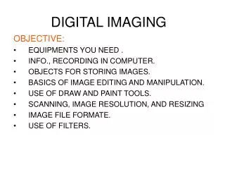
DIGITAL IMAGING. DIGITAL IMAGING. TERMINOLOGY. Film digitizer Digital radiography Digital fluoroscopy Dynamic range Matrix Pixel Imaging plate Histogram Algorithms Window level Window width TFT CCD. Post-processing image enhancement Latitude Analog Digital Image acquisition

E N D
TERMINOLOGY • Film digitizer • Digital radiography • Digital fluoroscopy • Dynamic range • Matrix • Pixel • Imaging plate • Histogram • Algorithms • Window level • Window width • TFT • CCD • Post-processing image enhancement • Latitude • Analog • Digital • Image acquisition • Image processing • Image display • Raster pattern • Laser • Photomultiplier tube • a-Selenium • ADC
SIMILARITIES AND DIFFERENCES FILM-SCREEN IMAGING DIGITAL & CR IMAGING • Need to select exposure factors • Accurate positioning • Use of accessory devices • IR receives radiation after passing thru patient • Latent image is produced and enhanced by the use of phosphorescence • Latent image is chemically processed • Limited dynamic range (30:1) • No post processing possible • Storage and retrieval issues • Need to select exposure factors • Accurate positioning • Use of accessory devices • IR receives radiation after passing thru patient • Latent image is produced (CR) and enhanced by the use of phosphorescence • Wide dynamic rage (1000:1) • Post Processing Enhancement is possible • Processing time reduced • Storage and retrieval easier
IMAGING PLATE Protective layer: Thin clear plastic that protects the phosphor layer Phosphor layer: this active layer contains the photo-stimuable-phosphor (barium fluorohalide phosphors) that react to x-ray exposure Reflective layer: reflects light forward when the plate is in the reader Conductive layer: absorbs the electrons released during exposure and reduces static electricity Color layer: absorbs stimulating light but reflects emitted light Support layer: semi- rigid layer that provides support Barcode label: allows technologist to use patient/exam identifying information
CR Phosphor Plates ABSORPTION EMISSION LASER STIMULATION ELECTRON TRAP ELECTRON TRAP X-RAY LIGHT
LATENT IMAGE (CR) • Formed by x-ray interaction with PSP • Ionizes phosphors, releasing electrons • Electrons trapped in crystal lattice of phosphor • Latent image is formed • Remains until processed by a reader • But does begin to decay so must be “read” in a timely fashion
IMAGE PROCESSING • PSP plate exposed to radiation • Electrons are trapped in phosphor layer • Plate is exposed to a red laser light • As electrons are released, a blue light is emitted • Blue light is captured and recorded by PMT • Image is sent to monitor for display • PSP plate is exposed to intense white light for erasure
DIRECT DIGITAL IMAGING Flat panel detector consists of plate covered with amorphous selenium (a-Selenium). This material absorbs x-rays and converts them to electrons. These electrons are stored in the TFT
TECHNIQUE CONSIDERATIONS • kVp Dependent, need mAs to saturate optimally • Now COMPUTER controls CONTRAST • Higher kVp to stimulate electron traps
80 kVp 200mAs 10 mAs 80 kVp Note Quantum Mottle
Histograms are used to plot density of data, and often for density estimation: estimating the probability density function of the underlying variable. The total area of a histogram used for probability density is always normalized to 1. If the length of the intervals on the x-axis are all 1, then a histogram is identical to a relative frequency plot. For x-ray purposes, a histogram tells how often a certain degree of gray is seen in the image.
To Produce Quality Images For Film/Screen Radiography or Digital/CR Radiography: The same rules, theories, and laws still apply and can not be overlookedSID, Inverse Square Law, Beam Alignment, Tube-Part-Film Alignment, Collimation, Grid, Exposure Factors: kVp, mAs, Patient Positioning
Quality Images Patient positioning • Accounts for 85% of the total number of repeat exposures. • Has a direct affect on exposure technique.
COLLIMATION CRITICAL • As the computer reads the density value of each pixel- it is averaged into the total • Close collimation= Better contrast • Bad collimation= more grays and less detail