Download

1 / 35
370 likes | 560 Views
The peripheral nerves. Dr. Faten. introduction. the nervous system contains many neurons + glial cells (neuroglia). Glial cells are very numerous; there are 10-50 times as many glial cells as neurons. The Schwann cells that invest axons in peripheral nerves are classified as glia .

E N D
The peripheral nerves Dr. Faten
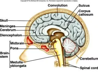
introduction • the nervous system contains many neurons + glial cells (neuroglia). • Glial cells are very numerous; there are 10-50 times as many glial cells as neurons. • The Schwann cells that invest axons in peripheral nerves are classified as glia. • In the CNS, there are three main types of neuroglia. 1-Microglia= (resemble tissue macrophages ) 2- Oligodendrogliocytes =are involved in myelin formation inside CNS. 3- Astrocytes=(form the blood-brain barrier & produce substances that are trophic to neurons) .
summary • NS Neurons + glial cells Cell bodies supporting cells Dendrites Schwann cells + Oligodendrogliocytes Axons (PNS) (CNS) (PNS) peripheral nerves conduct Impulses myelination myelination of CNS regeneration of PN
Definition of peripheral nerve • Axons of neurons with associated blood vessels and connective tissues are called peripheral nervesor simply nerves that carry sensory information into CNS and motor information from CNS. • An axon is a long cytoplasmic process capable of propagating an action potential
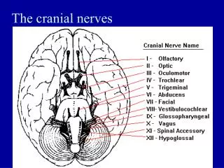
classification: Anatomical classification: 1. Cranial nerves: connected to the brain 2. Spinal nerves: attached to the spinal cord Functional Classification of nerves 1.Sensory = carry only sensory information 2.Motor = carry only motor information ( somatic, autonomic nerves). 3. Mixed = carry all. *Cranial nerves: some sensory; motor or mixed. * Spinal nerves are mixed nerves
Axon Diameter, mylination and conduction velocity =morphological and functional classification • Axons are classified into three groups according to the relationships among diameter, myelination, and propagation speed: 1. Type A fiber: largest axon (from 4 to 20 µm), myelinatedand its speed is 140 meters per second. 2. Type B fiber: Smaller (2-4 µm), myelinated axon, and its speed is 18 meters per second. 3. Type C fiber: Very small (less than 2 µm in diameter), unmyelinated and its speed is 1 meter per second. • We can understand the relative importance of myelin by noting that in going from Type C to Type A fibers, there is a tenfold increase in diameter, but the propagation speed increases by 140 times! For that the myelin increases the conduction speed.
The myelin • The myelin, a protein-lipid complex that is wrapped around the axon . • Outside the CNS, the myelin is produced by Schwann cells, glia-like cells found along the axon. • Myelin forms when a Schwann cell wraps its membrane around an axon up to 100 times. • The myelin sheath envelopes the axon except at its ending and at the nodes of Ranvier • The myelin has the insulating function. • The loss of myelin is associated with delayed or blocked conduction in the demyelinated diseases .
In the CNS , most neurons are myelinated, but the cells that form the myelin are oligodendrogliocytes rather than Schwann cells. • oligodendrogliocytes send off multiple processes that form myelin on many neighboring axons. • In multiple sclerosis, a crippling autoimmune disease, there is patchy destruction of myelin in the CNS.
Functions of each type Type A fiberscarry sensory information to the CNS concerning position, balance, and delicatetouch and pressure sensations from the surface of the skin. The motor neurons that control skeletal muscles also send their commands over large, myelinated Type A axons. Type B fibers and Type C fiberscarry less- urgent information concerningtemperature, pain, itching, and general touch and pressure sensations to the CNS and carry motor instructions to smooth muscle, cardiac muscle, glands, and other peripheral effectors.
NOTES * only around one-third of all axons carrying sensory information are myelinated, and most sensory information arrives over slender Type C fibers. * Myelination improves coordination and control by decreasing the time between reception of a sensation and initiation of an appropriate response. * Myelination begins relatively late in development, and the myelination of sensory and motor fibers is not completed until early adolescence.
Properties of nerves 1. Excitability=respond to various stimuli by electrical changes called action potential (nerve impulse). 2. Conductivity=the nerve can conduct impulses in both direction if stimulated at its middle. However in the body, the nerve conducts impulses in one direction only and this normal direction is called orthodromic conduction, but if it occurs in opposite direction is called antidromic direction ( example = triple response). *myelinated fibers used saltatory or jumping conduction ( ↑ velocity) from ranvier node to next. *non-myelinated fibers used continuous conduction (slowly process). (Review genesis of resting membrane and AP and propagation).
Saltatory Conduction • myelin is an effective insulator . • depolarization in myelinated axons jumps from one node of Ranvier to the next . • This jumping of depolarization from node to node is called saltatory conduction. • It is a rapid process, and myelinated axons conduct up to 50 times faster than the fastest unmyelinated fibers.
3. All or none =means that an increasing in stimulus intensity above threshold produce the same action potential with its threshold. 4. the various classes of fibers in peripheral nerves differ in their sensitivity to hypoxia and anesthetics. * Local anesthetics depress transmission in the group C fibers before they affect the touch fibers in the A group. *Conversely, pressure on a nerve can cause loss of conduction in large-diameter motor, touch, and pressure fibers while pain sensation remains relatively intact. *Patterns of this type are sometimes seen in individuals who sleep with their arms under their heads for long periods, causing compression of the nerves in the arms. *Because of the association of deep sleep with alcoholic intoxication, the syndrome is commonest on weekends and has acquired the interesting name Saturday night or Sunday morning paralysis.
Compound action potential: * Each nerve contains more than one type of nerve fibers with different speeds of conduction. *If an stimulus is applied at one point on a nerve and the action potential is recorded some distance away → several peaks will appear and represent the arrival of action potential in the fastest fibers then in the slower fibers. *This action potential with multiple peaks is called a compound action potential.
Neuronal response to nerve transection (cutting) 1. Degeneration and is usually followed by 2. Regeneration 1. Degeneration types A. wallerian degeneration B. retrograde degeneration C. transneural degeneration
degeneration A. wallerian (orthograde) degeneration= degenerative changes occur in the distal part of a nerve which is separated from their cell bodies if it is transected: *Swelling of neurofibrils and break into short segment *myelin hydrolysis beaked into oil droplets *all remnants are removed by the neurilemmal cells and tissue macrophages in the area. *neurilemmal tube becomes empty
B. retrograde degeneration ofproximal part and cell body: *degeneration of proximal part = like distal part changes * Degeneration of cell body = swelling, eccentric nucleus, fragmentation of Golgi apparatus and chromatolysis C. Transneuronal degeneration: • Means degeneration of a neuron if it becomes functionless after degeneration of another neuron.
Regeneration *Cell body regenerationstarts 20 days after transection and before nerve fiber regeneration and complete in about 80 days *cell regains normal size, nucleus returns to center and Golgi apparatus are restored. *nerve fiber regeneration starts by growing of proximal neurilemma tube till its cut ends. Then, it sprouts into several branches , at the same time the distal part grows towards sprouting neurofibrils by chemotaxis secreted by schwann cells. *The myelin sheath is regenerated by neurilemmal cells *schwann cells also release growth factors ( neurotrophins) such as nerve growth factor and insulin like growth factor to promote growth of nerve. -
Regeneration of a nerve after trauma • Tissues Macrophages migrates into trauma area • Phagocyize disintegrated myelin & axonal debris +release mitosis stimulating chemicals stimulate • Proliferation of shawnn cells by neurilemmal sheat that form cords to guide regeneration of axon & Release • Growth factors Stimulate Axonal growth Sprouting across gap to reach original contact
Notes : • Rate of nerve growth = 1mm / day (as maximum) • inside CNS, nerve fibers have no neurilemmal sheath so, after inj. no regeneration occurs. • neurilemmal sheath = schwann cells surrounded by basement membrane, then form a tube facilitates regeneration of nerve. But in CNS this continuous basement membrane not present , so no tube for regeneration. • unmyelinated nerve are also surrounded by a sheath of schwann, but it lacks the multiple wrappings of schwann cell membrane.