Download
1 / 78
780 likes | 789 Views
ORGANELLE-SPECIFIC PROTEIN QUALITY CONTROL SYSTEMS AND PROTEIN MISFOLDING DISEASES. Protein Folding and Quality Control Systems in the Cytosol. Comprises a large number of components. Upon emerging from the ribosome, nascent polypeptides are protected by chaperones, such a s

E N D
ORGANELLE-SPECIFICPROTEIN QUALITY CONTROLSYSTEMS AND PROTEINMISFOLDING DISEASES
Protein Folding and Quality ControlSystems in the Cytosol Comprises a large number of components. Upon emerging from the ribosome, nascent polypeptides are protected by chaperones, such as nascent-polypeptide-associated complex(NAC), Hsp40, Hsp70, prefoldin, and TCP-1 ring complex (TRiC), and held in a folding competent state until released from the ribosome.
Subsequently, most small proteins complete their folding in the cytosol without assistance, whereas a fraction of the cytosolic proteins requires further assistance from chaperones, e.g., Hsp90 and TRiC. TRiC, which is the most complex cytosolic chaperone, is composed of a double-ring structure, each with eight different subunits forming a large cavity in which the polypeptide is folded to a native or near-native form, and later released into the cytosol
If the folding to the native structure cannot be completed, the chaperones assess whether misfoldedconformers should be refolded or degraded by the ubiquitin-proteasome pathway in order to eliminate toxic conformations. Targeting a polypeptide for degradation requires a multistep pathway to covalently attach ubiquitin monomers.
Ubiquitin is activated by the ubiquitin-activating enzyme (E1), and transferred to an ubiquitin carrier protein (E2). The E2 enzyme and the polypeptide both bind to a specific ubiquitin protein ligase(E3), and ubiquitin is covalently attached to the substrate. Further steps generate a polyubiquitin chain, targeting the polypeptide substrate to the proteasome for degradation
The integrated system of chaperones and the components of the ubiquitin-proteasome pathway comprise the most important cytosolic PQC system. Failure of the PQC system to degrade misfolded proteins may lead to formation of protein aggregates. Aggregates in the cytosol may accumulate at a single site called an aggresome, or as soluble monomers and oligomers, which later may precipitate into long amyloid fibrils.
Aggresomes are large globular deposits formed by transport of aggregated material along microtubulartracks in a highly ordered transportation system, whereas amyloid fibrils are long protein aggregates with a tube-like core region formed by the inherent properties of circular β-sheet structures
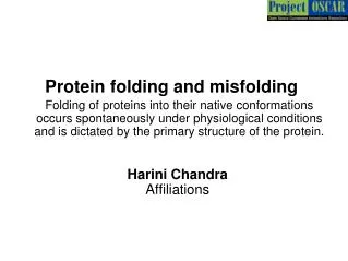
Cytosol-Associated ProteinMisfoldingDiseases (1) Defective folding caused by amino acid substitutions that result in rapid degradation of the variant protein is exemplified by phenylketonuria (PKU), an inborn error of phenylalanine metabolism. Amino acid substitutions in the phenylalanine hydroxylase (PAH) enzyme far from the enzyme’s active site may cause misfoldingof the protein, hinder formation of the active tetramer, and trigger rapid degradation.
In many cases PKU is thus due to loss-of-function pathogenesis. An increase in the chaperone concentration or lowering of the temperature facilitates the protein folding and, at least in cell models, rescues the enzymatic activity of PAH
Loss-of-functionpathogenesis: definition Pathogenesis resulting from insufficient protein functiondue to inadequate protein synthesis, functional site amino acid alterations, or inability to achieve the functional protein structure because of misfolding
PKU At A Glance • PKU is a metabolic disorder caused by a deficiency of the liver enzyme phenylalanine hydroxylase. It prevents normal metabolization of
PKU At A Glance • Phe, one of the essential amino acids that cannot be manufactured by the body and must therefore be taken from protein rich foods.
Phe to Tyr Conversion • Individuals with PKU have a deficiency in the enzyme
Phe to Tyr Conversion • phenylalanine hydroxylase, which converts phenylalanine to tyrosine.
Metabolic Pathways • In individuals with PKU, phenylalanine can’t be converted into tyrosine, and the metabolic process stops short of producing the needed end products.
Metabolic Pathways • Phenylalanine builds up in the body to toxic levels, causing mental retardation.
PKU Treatment • The only treatment available for PKU is a diet where phenylalanine levels are strictly limited.
PKU Prognosis • If the condition was not diagnosed early and a special diet started, the indidivudal will suffer severe and irreversable brain damage.
Cytosol-Associated ProteinMisfolding Diseases(2) A special case of protein misfoldingdiseases are those caused by variations in the folding machinery itself, leading to reduced PQC efficiency. One example is desmin-related myopathy, where αB-crystallin, a small heatshockprotein, plays a role in the folding of desmin, which has its function in the intermediate filaments of cardiomyocytes. This function, however, is compromised by a single amino acid substitution that leads to the formation of aggregates containing both desminand αB-crystallin
Molecular cytoarchitecture of a myocyte, featuring proteins involved in skeletal and cardiac myopathies. Desminis a main muscle protein. It interacts with other proteins to support myofibrils. Desmin provides maintenance of cellular integrity, force transmission, and mechano-chemical signaling. Mutations in other sarcomeric and cytoskeletal proteins (plectin,filaminC, αB-crystallinetc..) cause neuromuscular disorders
Cytosol-Associated ProteinMisfolding Diseases (3) In Parkinson’s disease (PD), protein aggregates are formed in the brain, leading to neurodegeneration. Point mutations or increased expression of the α-synuclein gene lead to a dominant form of the familial disease by a toxic gain-of-function pathogenesis due to cytosolic aggregates consisting of either wild-type or variant α-synuclein, as well as components of the ubiquitin-proteasome system.
Definition of Gain-of-function pathogenesis : The misfolded protein accumulates/ aggregates in the cell, giving rise to new toxic functions related to physico-chemical properties
The diversity of synucleinopathies overview of where different synucleinopathiesexist in brain Parkinson's disease is shown in orange and affects the substantianigra
Early onsetrecessive forms of Parkinson’s disease are associated with variations in the PARKIN, UCH-L1, DJ-1, or PINK1 genes. These genes code for components involved in the ubiquitinationand turnover of α-synuclein, and it is speculated that the pathogenesis includes a loss of PQC function, leading to α-synucleinaggregation in addition to general oxidative stress causing mitochondrial dysfunction
Cytosol-Associated ProteinMisfolding Diseases(4) The pathogenesis of amyotrophic lateral sclerosis (ALS) mediated by Cu,Zn superoxide dismutase gene (SOD1) variations is believed to be gain-of function through aggregation of the misfolded variant SOD1 protein. SOD1 protects the cell from oxidative damage by catalyzing the dismutation of the superoxide radicals to hydrogen peroxide and oxygen
Reaction catalyzed by SOD1 SOD1 catalyzes the dismutation of the superoxide radicals to hydrogen peroxide and oxygen The disease therefore seems to be a case of increased oxidative damagefrom enzymatic haploinsufficiency
However, artificial reduction of the enzymatic SOD1 activity does not mimic the ALS phenotype in animal models. In fact, several of the SOD1 variants remain fully active. More than 100 disease-associated SOD1 gene variations are known, accounting for 25% of the familial ALS cases, which for the most part are transmitted in a dominant fashion. Several gained functions have been proposed for the variant proteins, such as aberrant chemistry of the Cu and Zn sites, loss of protein function through co-aggregation with the aggregates, depletion of molecular chaperones, dysfunction of the proteasome overwhelmed with misfolded proteins, as well as disturbance of mitochondrion and peroxisome functions.
A combinedgain-of-function and loss-of function may be a more widespread pathogenesis than presently acknowledged, and a dysfunctional effect from accumulation of aberrant proteins may in fact be present in many protein misfolding diseases
Protein Folding and Quality Controlin the Endoplasmic Reticulum Endoplasmic reticulum (ER): first compartment of the secretory pathway. It is engaged with ribosomal protein synthesis, co- andpost-translational modification, and protein folding. Proteins enter the organelle in an unfolded state and begin to fold co-translationally.
ER lumen contains high concentrations of a specialized set of chaperones and folding enzymes, which assist protein folding in conjunction with post-translational modifications, e.g., signal peptide cleavage, disulfide bond formation, and N-linked glycosylation. In this respect, the ER plays a crucial role in the PQC, regulating the transport of proteins from the ER to the Golgi apparatus, as only proteins that have attained their native structure in the ER are exported efficiently
Interactions with components of the primary PQC, i.e., BiP, calnexin, calreticulin, glucose-regulated protein Grp94 and the thiol-disulphideoxidoreductases, protein disulphideisomerase (PDI), and ERp57assist protein folding. Misfoldedor unassembled proteins may accumulate in the absence of efficient ER associated degradation (ERAD)
Substrates for ERAD are selected by the PQC system and translocated to the cytosol, where they normally are degraded by the ubiquitin-proteasome system
A substantial number of cellular proteins are processed and transported through the ER. These include receptors and ion channels to be expressed on the cell surface, enzymes and hormones to be secreted, as well as proteins with a specialized function within the organelles of the secretory pathway. Because many of these proteins are essential and indispensable in many physiological processes, a variety of disease phenotypes may result from impairment of their ER-mediated transport. Therefore, defective ER processing of proteins may contribute to numerous diseases
Endoplasmic Reticulum–AssociatedMisfoldingDiseases (1) ER-associated misfolding and rapid degradation by ERAD are hallmarks of cystic fibrosis (CF), which is a lethal autosomal recessive disease caused by mutations in the CF transmembraneconductance regulator(CFTR ) gene encoding a chloride channel. The disease results from loss of chloride regulation in epithelia expressing the gene
More than 1000 disease-associated CFTR gene variations have been described. However, one single variation, coding for an one amino acid deletion variant, ΔF508, is the most common, accounting for about 66% of all disease- associated variant CFTR alleles worldwide. For the ΔF508 CFTR protein the maturation process is very inefficient and virtually all of the protein (>99%) undergoes rapid ERAD
CFTR Structure & Function
Cystic Fibrosis Transmembraneconductance Regulator (CFTR) is a protein that in humans is encoded by the CFTR gene. CFTR is an ABC transporter-class ion channel that transports chloride and thiocyanate ions across epithelial cell membranes. Structure:- CFTR is a glycoprotein with 1480 aa. It consists of five domains. There are two transmembrane domains, each with six spans of alpha helices. These are each connected to a nucleotide binding domain (NBD) in the cytoplasm. The first NBD is connected to the second transmembrane domain by a regulatory "R" domain that is a unique feature of CFTR, not present in other ABC transporters.
CFTR is composed of five functional domains. TMDs or ‘transmembrane domains’: Around 19% of CFTR is composed of TMD1 and TMD2, which form the channel pore allowing transport of chloride ions across the membrane. NBDs or ‘nucleotide-binding domains’: These domains bind the nucleotide molecule ATP (a vehicle of chemical energy). Opening and closing of the channel (or ‘gating’) requires ATP to bind to these domains. Regulatory (‘R’) domain: The R domain regulates channel activity and can be considered to be the ‘trigger’ governing whether the channel opens or closes, to activate the channel. Many CF-causing mutations occur in NBD1, including F508del,
The ion channel only opens when its R-domain has been phosphorylated by PKA and ATP is bound at the NBDs. The carboxyl terminal of the protein is anchored to the cytoskeleton by a PDZ-interacting domain**.
Function: CFTR functions as a -activated ATP- gated anion channel, increasing the conductance for certain anions (e.g. Cl–) to flow down their electrochemical gradient. ATP-driven conformational changes in CFTR open and close a gate to allow transmembrane flow of anions. The CFTR is found in the epithelial cells of many organs including the lung ,liver, pancreas, digestive tract, reproductive tract, and skin. Normally, the protein moves chloride and ions with a negative charge out of an epithelial cell to the covering mucus.
**The PDZ domain is a common structural domain of 80-90 amino-acids found in the signaling proteins of bacteria, yeast, plants, viruses and animals. Proteins containing PDZ domains play a key role in anchoring receptor proteins in the membrane to cytoskeletal components. PDZ domain structures. (A) PDZ3 of PSD-95 (cyan), complexed with the C-terminal pentapeptide of CRIPT (KQTSV, yellow). (B) The PDZ domain of a-1 syntrophin (green), complexed with the PDZ domain of nNOS (blue). (C)Homodimer of Grip1 PDZ6 (pink and purple), complexed with the C-terminal octapeptide of Liprin (ATVRTYSC, yellow).
Positively charged sodium ions follow these anions out of the cell to maintain electrical balance. This increases the total electrolyte concentration in the mucus, resulting in the movement of water out of cell by osmosis. Mutation Well over one thousand mutations have been described that can affect the CFTR gene. Such mutations can cause two genetic disorders, congenital bilateral absence of vas deferens and the more widely known disorder cystic fibrosis. Both disorders arise from the blockage of the movement of ions and, therefore, water into and out of cells. In congenital bilateral absence of vas deferens, the protein may be still functional but not at normal efficiency, this leads to the production of thick mucus, which blocks the developing vas deferens.
In people with mutations giving rise to cystic fibrosis, the blockage in ion transport occurs in epithelial cells that line the passage ways of the lungs, pancreas, and other organs. This leads to chronic dysfunction, disability, and a reduced life expectancy. The most common mutation, ΔF508 results from a deletion (Δ) of three nucleotides which results in a loss of the amino acid phenylalanine (F) at the 508th position on the protein. As a result the protein does not fold normally and is more quickly degraded.
Synthesis of CFTR occurs with its concomitant insertion in the ER membrane and attachment of Hsc70/Hsp70 to nascent cytosolic domains. The cell seems to use this Hsc70/Hsp70 control as the first checkpoint to assess CFTR conformation, and it has been proposed that it is the major mechanism to discard F508 CFTR. In contrast, wild-type CFTR proceeds in the folding pathway through interaction of its N-glycosyl residues with calnexin. Subsequently, it acquires its native conformation for ER export through successive rounds of de- and re-glucosylation binding to calnexin.
The cellular fate of CFTR chloride channels is depicted. Wild-type CFTR is transported to the plasma membrane. By contrast, ΔF508-CFTR, the mutant protein present in individuals with cystic fibrosis, is degraded by endoplasmic-reticulum-mediated pathways (ERAD) before it reaches the plasma membrane.
Endoplasmic Reticulum–AssociatedMisfoldingDiseases (2) Like CF, hereditary emphysema dueto α-1-antitrypsin deficiency seems to involve ER associatedmisfoldingand rapid degradation. α-1-antitrypsin is a major plasma serine protease inhibitor secreted by hepatocytes to regulate the proteolyticactivity of various circulating enzymes. α-1-antitrypsin shows considerable genetic variability, having more than 90 naturally occurring variants