Download
1 / 30
300 likes | 485 Views
Case Report Diabetic artefacts in forensic practice. O.P. Murty MBBS MD (Professor) * Forensic Medicine, Faculty of Medicine, UITM, Malaysia 40450 Present by.Cap.Sudhunsa Suebsaman. Diabetic artefacts in forensic practice.

E N D
Case ReportDiabetic artefacts in forensic practice O.P. Murty MBBS MD (Professor) * Forensic Medicine, Faculty of Medicine, UITM, Malaysia 40450 Present by.Cap.Sudhunsa Suebsaman
Diabetic artefacts in forensic practice... • A case is presented where confusion arose about skin lesions and whether they were diabetic or electricalin origin. The deceased was a known diabetic and hypertensive man. A middle-aged person in early fifties was found unconscious in the cell and judicial autopsy was performed.He was facing trial for capital punishment of being allegedly involved in drug trafficking and money laundering. He had few marks over his fingers and foot which were considered to be electric marks produced in electric torture.
Electrical injuries - Electrical injuries can cause multiorgan dysfunction and a variety of burns and traumatic injuries 1.Cardiovascular : arrhythmia, asystole
Electrical injuries • Respiratory: respiratory arrest • Musculoskeletal: compartment syndrom • ENT/head: perforated tympanic membranes, facial burn,
Electrical injuries • Skin: A variety of burns and thermal injuries occurs from electricity that affect the skin and soft tissues
Electrical injuries High-voltage electrothermal burns: - Contact point
Electrical injuries High-voltage electrothermal burns - Ground point
Electrical injuries • Low-voltage burns: erythema to full-thickness burns.
Diabetes mellitus • Is a condition in which the body either does not produce enough, or does not properly respond to, insulin, a hormone produced in the pancreas. Insulin enables cells to absorb glucose in order to turn it into energy. This causes glucose to accumulate in the blood (hyperglycemia), leading to various potential complications.
Diabetes mellitusComplications : 1. Microangiopathy - Diabetic cardiomyopathy : heart failure -Diabetic nephropathy : kidney failure - Diabetic neuropathy :muscle weakness due to neuropathy , DM foot ,Sensory neuropathy
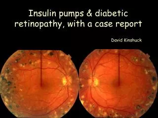
Diabetes mellitus • Stroke : mainly the ischemic type • Diabetic retinopathy : Vision loss , Blindness • Peripheral vascular disease, - periungual erythema
Diabetes mellitus • Dermopathy - Spontaneous Blisters in Diabetes
Case summary • The deceased was a middle-aged under trial and was lodged in state jail. He was a known diabetic and hypertensive man. He had fallen on the floor inside the room in prison and became unconscious.He was taken to prison hospital and from there immediately taken to district hospital. CT scan showed, fracture of skull bones, extradural haemorrhage,subdural Haemorrhage, cerebral oedema,brain infarction on right side. He underwent craniotomy twice for drainage of blood which had collected due to stroke and head injury.
Fig. 1. Solitary-confinement cell Fig. 2. Place of fall in the cell where he sustained fracture of skull in occipital area after stroke, CVA.
External examination Fig. 3. Middle occipital areas showed abraded bruise near cut hair.
External examination Fig. 11. Hand showing necrotic nail bed areas. Fig. 15. Finger nail beds are clean without any ischemic effect.
External examination Fig. 12. Enlarged and close-view of above areas. Blackish discoloration of nail beds,right index finger with a large blackish-blue area ; ring finer just showing early appearance of start of discolorations ; ring finger showing eroded necrotic ulcerative lesion ; little finger nail also showing small eroded ulcerative lesion.
External examination Fig. 13. Foot showing reddish areas (A and B).
External examination Fig. 14. Shows early erythematic blister like appearances in toes except the great toe Fig. 16. Foot of other side in clean.
Internal examination • Central nervous system Fig. 3. Middle occipital areas showed abraded bruise near cut hair.
Internal examination Central nervous system Fig. 4. Blood in occitpal and tempero-parietal area of scalp, exaggerated by craniotomy wound Fig. 6. Suturing of fracture line with craniotomy holes.
Internal examination Fig. 7. Under surface of brain with contusion haemorrhages. Atherosclerosis of vessels also visible. Fig. 8. Necrotic brain tissue after CVA.
Internal examination • Cardiovascular system Fig. 10. Advance atherosclerosis in descending and abdominal aorta
Discussion • In our study the lesions seen in the finger beds and soles could be due to diabetes in this deceased. But main concern is as how to deny allegations in this case of whether he was tortured electrically in the prison as lesions are closely simulating the finer electric points which are seen in the victims tortured in the jail.
Discussion Artefacts Fig. 12. Enlarged and close-view of above areas. Blackish discoloration of nail beds,right index finger with a large blackish-blue area ; ring finer just showing early appearance of start of discolorations ; ring finger showing eroded necrotic ulcerative lesion ; little finger nail also showing small eroded ulcerative lesion. Fig 21. Finger of a patient with diabetes mellitus demonstrating erythema of the proximal nail fold. This erythema is associated with dilatation of the superficial vascular plexus.
Discussion Artefacts Electrical injuries Ground point Fig. 14. Shows early erythematic blister like appearances in toes except the great toe
Discussion • In this particular case skin lesions were not seen when he was admitted in the hospital. He remained in coma for few days and after that he developed chest infection.The lesions were noticed only just two day prior to his death. • His autopsy report mentioned that he was a case of accidental fall after stroke from a standing height in the solitary confinement prison cell.
Discussion • The lesions over the skin of hand and feet were probably due ischemic effects because of poor neuro-muscular and vascular control as a result of cerebral ischemia Spontaneous Blistersin Diabetes periungual erythema
Discussion • Opinion: death was due to cerebral hypoxia consequent to brain infarction and head injury (a) Cranio-cerebral injury Due to (b) Fall from standing height Due to (c) Brain infarction
Do you have any question ? Thank you.....Ka