Download
1 / 1
10 likes | 169 Views
* corresponding author : karasd@seznam.cz. 1 Department of Medical Chemistry and Biochemistry, Palacký University, Hněvotínská 3, CZ-775 15 Olomouc, Czech Republic, 2 Institute of Microbiology AS CR, Vídeňská 1083, Prague 4, CZ 142 20, Czech Republic ,

E N D
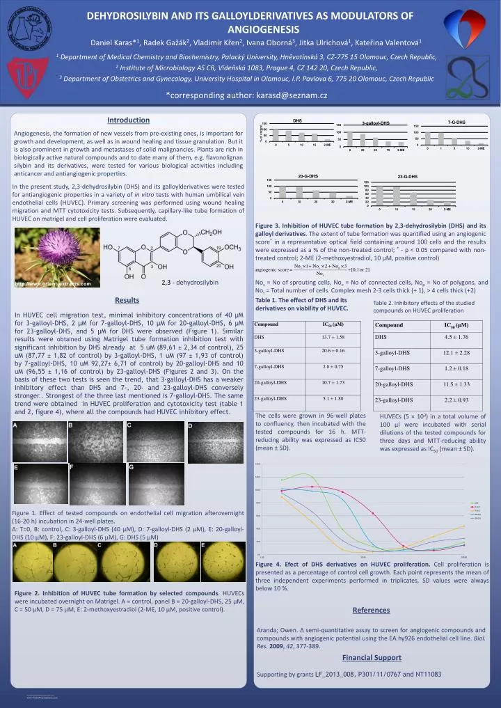
*correspondingauthor: karasd@seznam.cz 1 Department of Medical Chemistry and Biochemistry, Palacký University, Hněvotínská 3, CZ-775 15 Olomouc, Czech Republic, 2 Institute of Microbiology AS CR, Vídeňská 1083, Prague 4, CZ 142 20, Czech Republic, 3 Department of Obstetrics and Gynecology, University Hospital in Olomouc, I.P. Pavlova 6, 775 20 Olomouc, Czech Republic DEHYDROSILYBIN AND ITS GALLOYLDERIVATIVES AS MODULATORS OF ANGIOGENESIS Daniel Karas*1, Radek Gažák2, Vladimír Křen2, Ivana Oborná3, Jitka Ulrichová1, Kateřina Valentová1 Supporting by grantsLF_2013_008, P301/11/0767 and NT11083 Financial Support In HUVEC cell migration test, minimal inhibitory concentrations of 40 µM for 3-galloyl-DHS, 2 µM for 7-galloyl-DHS, 10 µM for 20-galloyl-DHS, 6 µM for 23-galloyl-DHS, and 5 µM for DHS were observed (Figure 1). Similar results were obtained using Matrigel tube formation inhibition test with significant inhibition by DHS already at 5 uM (89,61 ± 2,34 ofcontrol), 25 uM (87,77 ± 1,82 ofcontrol) by 3-galloyl-DHS, 1 uM (97 ± 1,93 ofcontrol) by 7-galloyl-DHS, 10 uM 92,27± 6,71 ofcontrol) by 20-galloyl-DHS and 10 uM (96,55 ± 1,16 ofcontrol) by 23-galloyl-DHS (Figures 2 and 3). On the basis of these twotestsisseenthe trend, that 3-galloyl-DHS has a weaker inhibitory effectthan DHS and 7-, 20- and 23-galloyl-DHS converselystronger.. Strongestofthethree last mentionedis 7-galloyl-DHS. Thesame trend wereobtained in HUVEC proliferationand cytotoxicity test (table 1 and2, figure 4), whereallthecompounds had HUVEC inhibitory effect. References Results Introduction Angiogenesis, the formation of new vessels from pre-existing ones, is important for growth and development, as well as in wound healing and tissue granulation. But it is also prominent in growth and metastases of solid malignancies. Plants are rich in biologically active natural compounds and to date many of them, e.g. flavonolignan silybin and its derivatives, were tested for various biological activities including anticancer and antiangiogenic properties. Aranda; Owen. A semi-quantitative assay to screen for angiogenic compounds and compounds with angiogenic potential using the EA.hy926 endothelial cell line. Biol. Res.2009, 42, 377-389. In the present study, 2,3-dehydrosilybin (DHS) and its galloylderivatives were tested for antiangiogenic properties in a variety of in vitro tests with human umbilical vein endothelial cells (HUVEC). Primary screening was performed using wound healing migration and MTT cytotoxicity tests. Subsequently, capillary-like tube formation of HUVEC on matrigel and cell proliferation were evaluated. Figure 3. Inhibition of HUVEC tube formation by 2,3-dehydrosilybin (DHS) anditsgalloylderivatives. The extent of tube formation was quantified using an angiogenic score* in a representative optical field containing around 100 cells and the results were expressed as a % of the non-treated control; * - p < 0.05 compared with non-treated control; 2-ME (2-methoxyestradiol, 10 µM, positive control) 2,3 - dehydrosilybin Nos = No of sprouting cells, Noc = No of connected cells, Nop = No of polygons, and Not = Total number of cells. Complex mesh 2-3 cellsthick (+ 1), > 4 cellsthick (+2) Table 1. Theeffectof DHS anditsderivatives on viabilityof HUVEC. Table 2. Inhibitory effectsofthestudied compounds on HUVEC proliferation Thecellsweregrown in 96-wellplates to confluency, thenincubatedwiththetestedcompoundsfor 16 h. MTT-reducingabilitywasexpressed as IC50 (mean ± SD). HUVECs (5 × 10³) in a total volume of 100 µl wereincubatedwithserialdilutionsofthetestedcompoundsforthreedaysand MTT-reducingabilitywasexpressed as IC50 (mean ± SD). Figure 1. Effectoftestedcompounds on endothelial cell migrationafterovernight (16-20 h) incubation in 24-wellplates. A: T=0, B: control, C: 3-galloyl-DHS (40 μM), D: 7-galloyl-DHS (2 μM), E: 20-galloyl-DHS (10 μM), F: 23-galloyl-DHS (6 μM), G: DHS (5 μM) Figure 4. Efectof DHS derivatives on HUVEC proliferation. Cell proliferationispresented as a percentageofcontrol cell growth. Each point representsthemeanofthree independent experimentsperformed in triplicates, SD valueswerealwaysbelow 10 %. Figure 2. Inhibition of HUVEC tube formation byselectedcompounds. HUVECs were incubated overnight on Matrigel. A = control, panel B = 20-galloyl-DHS, 25 µM, C = 50 µM, D = 75 µM, E: 2-methoxyestradiol (2-ME, 10 µM, positive control).


