Download
1 / 56
560 likes | 681 Views
Chapter 15. The Chromosomal Basis of Inheritance. TOPICS FOR TODAY’S LECTURE. Morgan & Strutevant Sex Chromosomes Linked Genes Gene Map Why you are smarter than Sarah Palin Human Genetic Disorders. Chromosome theory of inheritance supported Mendel’s laws. (what are mendel’s laws?) 1.

E N D
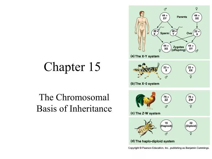
Chapter 15 The Chromosomal Basis of Inheritance
TOPICS FOR TODAY’S LECTURE • Morgan & Strutevant • Sex Chromosomes • Linked Genes • Gene Map • Why you are smarter than Sarah Palin • Human Genetic Disorders
Chromosome theory of inheritance supported Mendel’s laws (what are mendel’s laws?) 1. 2.
Chromosome theory of inheritance supported Mendel’s laws 1. Law of Segregation- pairs of factors separate during gamete formation 2. Law of Independent Assortment- separated pairs of factors sort themselves into gametes independently of each other.
THOMAS HUNT MORGAN was the first scientist to associate a specific gene with a specific chromosome. His experiments provided convincing evidence that chromosomes are the location of Mendel’s heritable factors. Gene stained with fluorescent dye shows the same locus on homologous chromosomes.
Experimental organism = Fruit fly = Drosophila melanogaster
Discovery of Sex Linkage(sex experiments w/ flies = creepy) • Cross #1 : • P generation • (pure breeding) • F1 results: all had red eyes=wild type • Suggests: dominance • like Mendel’s experiment red-eyed (wild-type) female white-eyed (mutant) male X
CROSS #2 (bro x sis) F1 x F1 = F2 F2 results: • 3 wild type: 1 mutant • Except that: • only males are mutant • all the females are wild type and • Males are mutant 1/2 of the time.
Red is wild type White is mutant… F1- all are wild type F2- all of the females are wild type. 1/2 males are wild type Discovered Sex chromosomes The gene for eye color was Inherited differently among Male and female flies in the F2. Difference: x & y chromosomes Genes located on a sex chromosome Are called X LINKED. Or Y LINKED Figure 15.3 Sex-linked inheritance
GENE LINKAGE & MAPS The number of genes in a cell is far greater than the number of chromosomes; in fact, each chromosome has hundreds or thousands of genes. Thus, linked genes tend to be inherited together.
Gene Linkage and Map Units • Gene linkage was explained by Thomas Hunt Morgan in 1910. • When he was examining traits of the fruit fly- • Drosophila melanogaster. Why study the fruit fly? Great for research because it was benign (harmless) unlike MEDFLY Reproductive cycle: 2 weeks Small genome: 2n is 8 -so great for genetic research.
Gene Linkage and Map Units • Genes are said to be LINKED when they • exist on the same chromosome • and they are inherited together. The genes of chromosome 9 are “LINKED”.
Gene Linkage and Map Units • When genes are linked, the expected phenotype ratios during breeding experimentsdeviate from the Mendelian ratios of: 1) F1 xF1 dihybrid cross Ex (AaBb x AaBb) F2 = 9:3:3:1 More importantly: 2) F1 dihybrid test cross Ex (AaBb x aabb) F2 = 1:1:1:1
For Example: • Parents who were pure for two traits (homozygous) were crossed (AABB x aabb). • The F1 generation produced individuals that were heterozygous for both traits. (AaBb) • An F1 individual is test-crossed with a homozygous recessive individual. (AaBb x aabb) • If both genes were located on different chromosomes the expected phenotypic ratio should be 1:1:1:1. • The actual ratios suggested that the genes DID NOT assort independently. Most of the phenotypes matched the P1 generation parents.
Recombinant phenotypes (those different from either P1 gen parents) were the result of crossing over instead of independent assortment.
The recombination frequency can be calculated by dividing the total number of recombinants by the total number of offspring.
One of Morgan’s students, Alfred Sturtevant came up with a method for constructing a genetic map, showing the position of genes on a chromosome. • An important observation the recombination frequencies reflect the distances between genes. • Therefore, genes farther apart have a greater chance of being separated by crossing over. • As the distance between genes increases, so does their recombination frequency. • The distance between genes are expressed in “map units” where one map unit = 1% recombination frequency. • Map units are called centimorgans, in honor of Morgan.
Morgan’s work: Evidence of “linked genes” in drosophila. Evidence of the chromosomal basis of inheritance.
WHY YOU ARE SMARTER THAN SARAH PALIN… “Where does a lot of that earmark money end up anyway? […] You've heard about some of these pet Projects… they really don't make a whole lot of sense and sometimes these dollars go to projects that have little or nothing to do with the public good. Things like fruit fly research in Paris, France. I kid you not.” Palin "fruit fly research"
Next- human genetic disorders. http://www.youtube.com/watch?v=Eg1vIeuQT1s
What is the difference between an infectious disease and a genetic one??? • Infectious illnesses/ diseases caused by microorganisms: virus, bacteria, protists, fungi, tiny animals (worms) that harm cells. • They can be transmitted from one host to another… contagious. • Genetic is inherited. Defect in the DNA code.
GENETIC DISEASES/DISORDERS • Thousands of genetic disorders are caused by recessive genes (mutations in DNA). • Some are mild and some are deadly. • Most of these alleles code for a malformed protein or for creating no protein at all. • NOT CONTAGIOUS, but can be transmitted to offspring.
Inheritance Patterns of Genetic Disorders: • Autosomal Recessive gg = disease • Autosomal Dominant Gg = disease • Autosomal Codominant • Sex Linked Recessive XgXg XgY = disease • Sex Linked Dominant XGXg XGY = disease • ANEUPLOIDY of autosomes (too many/few) disease • ANEUPLOIDY of sex chromosomes= “ “ • CHROMOSOME alteration during crossing over
Examples of recessively inherited disorders (autosomal recessive) • Cystic fibrosis: most lethal disease in the US Caused by defective chloride membrane channels. Leads to thick mucus building up in the lungs, digestive tract… low weight, susceptible to respiratory infections. 2) Tay-Sachs: caused by a dysfunctional enzyme that no longer breaks down fats. Leads to fatty build-up in the brain and around nerves. Accumulation of the lipids in brain cells causes progressive nervous system dysfunction and is usually fatal by age four.
3) Phenylketonuria (PKU): inability to properly break down the amino acid phenylalanine. Untreated causes mental retardation. Managed with restricted diet low in phenylalanine.
Ex. AUTOSOMAL CODOMINANT disorder: Sickle Cell: caused by a single amino acid substitution in hemoglobin. Red blood cells of individuals with this defect are unable to effectively transport oxygen throughout the body. (Pleiotropic effect on multiple organs)
Ex. AUTOSOMAL DOMINANT disorders: • Huntington’s disease: is a degenerative disease of the nervous system, the allele expresses itself later in life so although it is caused by a single allele and is lethal, it may already be passed on to the next generation. • Achondroplasia (dwarfism): Characterized by problems with bone growth. Also… polydactyl
Sex Linked Disorders & Patterns of Inheritance NO heterozygote among males because the gene is on the X chromosome… males are XY. Heterozygous females are “carriers”. Sex linked recessive: Males more susceptible to disease. 1. ALD (adreno leuko dystrophy)- sex linked recessive 2. Red-Green Color Deficiency- sex linked recessive 3. Hemophilia- sex linked recessive Sex linked dominant: Females more susceptible to disease. 1.Duchenne’s Muscular Dystrophy- sex linked DOMINANT
In humans, such X linked inheritanceIs designated XCXC XCXc XCXC XcY XCY Ishihara color blindness test Red Green Color Blindness is much more prevalent among males.
Hemophilia- inability to code for all factors required to form normal blood clots. Surface wounds ok. • Duchenne’s Muscular Dystrophy- Absence of an essential muscle protein. Results in deteriorating muscles and loss of coordination. (sex linked dominant- so females are more likely to show the disorder than other x-linked ones)
HEMOPHELIA Large hemorrhage… surface wounds are slow to heal but not Fatal- it is the internal bleeding and tissue wounds that are a problem
EUROPEAN ROYALTY AND HEMOPHILIA • History's most famous carrier of the gene for hemophilia was Victoria (1819-1901), Queen of England and grandmother to most of the royalty in Europe. In 1853, Queen Victoria gave birth to her eighth child, Leopold, Duke of Albany, who had hemophilia and died at the age of 31 from internal bleeding after a fall. • Two of Queen Victoria's four daughters, Alice (b. 1843) and Beatrice (b. 1857), also carried the gene for hemophilia and subsequently transmitted the disease to three of Victoria's grandsons and to six of her great-grandsons. • Alice's daughter Alexandra also was a carrier of hemophilia, and she transmitted the disease to her son Alexis (b. 1904), whose father was Czar Nicholas 11 (1868—1918) of Russia. Alexis is perhaps the most famous of the European royals with hemophilia. Alexis was the heir to his father's throne and his medical condition caused much anxiety in the royal household. Historians are still discussing the role Alexis's condition played in the Russian revolution of 1918.
Autosomal chromosome disorders • If an organism is born with an abnormal number of chromosomes it is called aneuploidy. • It is caused by nondisjunction of homologous chromosomes during meiosis. • The chromosomes fail to separate and one gamete receives both copies and the other gamete receives none.
Down syndrome or Trisomy 21 is caused by having three copies of the 21st chromosome.
Sex Chromosome disorders: • X inactivation: during embryonic development in females one X chromosome randomly condenses into an inactive mass (called a Barr body) within each cell… so each cell has only one active X chromosome. • The result: most of the alleles on the X chromosome are expressed individually. • X-inactivation, is an epigenetic change that results in a different phenotype but is not a change at the genotypic level. • This can give rise to mild symptoms in female ‘carriers’ of X-linked genetic disorders. • Reversed in the female germline, so that all oocytes contain an active x chromosome
In cats: this leads to tortoiseshell coloration because in • Some cells one x is inactive and in others, the other is. • In humans: females will express recessive disease alleles • more frequently… EX: faulty sweat glands in some areas.
2) Nondisjunction of SEX CHROMOSOMES a)XXY individuals: Klinefelter syndrome • Male sex organs • Small, sterile testes • Female body characteristics, including some breast development
2) Nondisjunction of SEX CHROMOSOMES a)XXY individuals: Klinefelter syndrome • Male sex organs • Small, sterile testes • Female body characteristics, including some breast development • XYY individuals: no associated syndrome • Taller than average c) XXX individuals: super females (trisomy x) taller than average, slightly lower intelligence • XO individuals: Turner syndrome • Sterile • Sex organs do not mature
3) Altering Chromosome Structure= usually occurs during cell division/ chromosome replication. • Deletion: removing a segment of chromosome ex. Cri du chat syndrome • Duplication: segments on a chromosome are repeated • Inversion: sections of the chromosome are reversed • Translocation: a segment of one chromosome is broken off and reattached on another non-homologous chromosome.
GENOMIC IMPRINTING Genetic phenomenon by which certain genes are expressed in a “parent-of-origin-specific ” manner. Inheritance process independent of classical Mendelian Inheritance. Involves “methylation” or silencing of genes as well as histone activation of others. VIDEO: “GHOST IN YOUR GENES”