Download
1 / 37
380 likes | 772 Views
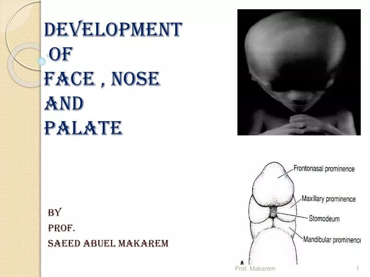
DEVELOPMENT OF FACE , NOSE AND PALATE. By Prof. Saeed Abuel Makarem. Development The of Face. The facial primordia appear early in the fourth week around the primordial stomodeum. FN. MX. MX. md. md. S. Five facial primordia appear as prominences around the stomodeum

E N D
DEVELOPMENT OF FACE , NOSE AND PALATE By Prof. Saeed Abuel Makarem Prof. Makarem
Development The of Face • The facial primordia appear early in the fourth week around the primordial stomodeum FN MX MX md md S Prof. Makarem
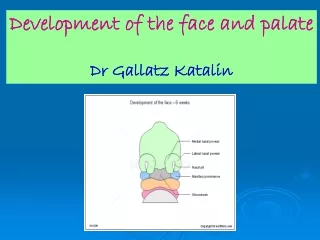
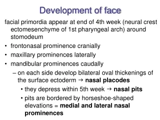
Five facial primordia appear as prominences around the stomodeum • The single frontonasal prominence • The paired maxillary prominences • The paired mandibular prominences FNP Prof. Makarem
The paired facial prominences are derivatives of the first pair of pharyngeal arches • The prominences are produced mainly by the proliferation of neural crest cells. • These cells migrate from the Mesencephalonand & Rhombencephalon regions of the neural folds into the arches during the fourth week 1st Prof. Makarem
The Nasal placodes: • Two bilateral oval thickenings in the surface ectoderm, of the frontonasal prominence, appear, by the end of the fourth week. • Nasal placodes are the primordia of the nose and nasal cavities. Initially these placodes are convex. • Later, they are stretched to produce a flat depression in each placode. Prof. Makarem
Mesenchyme in the margins of the placodes proliferate, producing horse shoe shaped elevations called medial and lateral nasal prominences • Now the nasal placodes lie in depression called nasal pits • These pits are the primordia of the anterior nares (nostrils) and nasal cavities Prof. Makarem
Mesenchymal cells are the major source of the connective tissue components, including muscles,cartilage, bone, and ligamentsin the facial and in the oral regions. Prof. Makarem
The frontonasal prominence (FNP) is formed of 2 parts: • 1- Frontal part: forms the forehead • 2- Nasal part: forms the rostral boundary of the stomodeum. • The paired maxillary prominences form the lateral boundaries of the stomodeum • The paired mandibular prominences constitute the caudal boundary of the primitive mouth Prof. Makarem
Facial development occurs mainly between the fourth and eighth weeks. • By the end of the embryonic period, (8th week) the face has an unquestionabl human appearance. Prof. Makarem
Between the 7th and 8th weeks, the two medial nasal prominences merge with each other and with the maxillary and lateral nasal prominences Merging of the medial nasal and maxillary prominences results in continuity of the upper jaw and upper lip and separation of the nasal pits from the stomodeum Prof. Makarem
The lower jaw and lower lips are the first parts of the face to form • They result from merging of the medial ends of the mandibular prominences in the median plane • Median cleft lower lip is a very rare condition M Prof. Makarem
Each lateral nasal prominence is separated from the maxillary prominence by a cleft called nasolacrimal groove Prof. Makarem
The nasolacrimal duct develops from a rodlike thickening of ectoderm in the floor of the nasolacrimal groove • This thickening gives rise to a solid epithelial cord that separates from the ectoderm and sinks into the mesenchyme • As a result of cell degeneration, this epithelial cord canalizes to form the nasolacrimal duct Prof. Makarem
The cranial end of this duct expands to form the lacrimal sac • By the late fetal period, the nasolacrimal duct drains into the inferior meatus in the lateral wall of the nasal cavity • The duct usually becomes completely patent only after birth • Occasionally, part of the duct fails to canalize causing atresia of the nasolacrimal duct. Prof. Makarem
Development of the External Ear • By the end of the fifth week, the primordia of the auricles of the ears have begun to develop • Six auricular hillocks form around the first pharyngeal groove (cleft). • Three on each side of the 1st pharyngeal groove (cleft). • These are the primordia of the auricle and external acoustic meatus. • Initially the ear located in the neck. • As the mandible develops the ears ascend to the level of the eye. Prof. Makarem
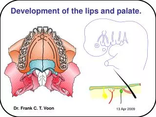
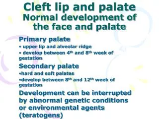
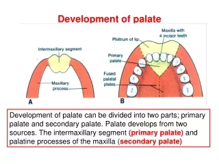
Development of The Palate As the medial nasal prominences merge, they form an intermaxillary segment The intermaxillary segment gives rise to: 1- The Philtrum(median part of the upper lip). 2- The Premaxillary part of the maxilla and associated gingiva (gum). 3- The primary palate. Prof. Makarem
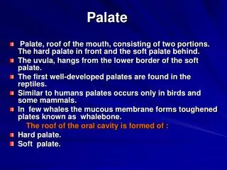
Development of Palate The palate develops from two stages: • Primary palate • Secondary palate • Palatogenesis begins at the end of the fifth week and is completed at twelfth week (5------12 ) The critical period of the palate development is from the end of the sixth week until the beginning of ninth week (6 ------ 9) Prof. Makarem
Primary Palate • Early in the sixth week the primary palate or median palatine process begins to develop from the intermaxillary segment of the maxilla • Initially this segment is formed by merging of the medial nasal prominences • The primary palate forms the premaxillary part of the maxilla • It represents only a small part of the adult hard palate • (2 Medial nasal prominences >>>>intermaxillary segment .>>>>>>>>>-primary palate). Prof. Makarem
Secondary Palate • The secondary palate is the primordium of the hard and soft palate • It begins to develop early in the sixth week from two mesenchymal projections that extend from the internal aspects of the maxillary prominences Prof. Makarem
Initially the lateral palatine processes or palatalshelves project inferomedially on each side of the developing tongue Prof. Makarem
As the jaws develop, the tongue becomes relatively smaller and moves inferiorly • During the 7th & 8th weeks, the lateral palatine processes elongate and ascend to a horizontal position superior to the tongue • Gradually these processes approach each other and fuse in the median plane Prof. Makarem
Palatine processes also fuse with the nasal septum and the posterior part of the primary palate • The nasal septum develops as a downgrowth from internal parts of the merged medial nasal prominences • The fusion between the nasal septum and the palatine processes begins anteriorly during the ninth week and is completed posteriorly by the twelfth week Prof. Makarem
Palatal shelves move medially and fuse with the nasal septum. Prof. Makarem
Bone gradually develops in the primary palate, forming the premaxillary part of the maxilla, which lodges the incisor teeth • Concurrently bone extends from the maxillae and palatine bones into the lateral palatine processes to form the hard palate • The posterior part of these processes do not ossified. Prof. Makarem
They extend posteriorly beyond the nasal septum and fuse to form the soft palate. • Its soft conical projection is called uvula • The median palatine raphe indicates the line of fusion of the lateral palatine processes Prof. Makarem
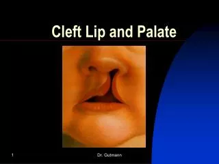
Cleft Lip and Palate • The defect is usually classified according to developmental criteria There are two major groups of cleft lip and palate: • Clefts involving the upper lip and anterior part of the maxilla • Clefts involving the hard and soft regions of the palate Prof. Makarem